Nachawi Noura, Lew Madelyn, Konopka Kristine, Sandouk Zahrae
1Department of Internal Medicine, Saint Joseph Mercy Hospital Ann Arbor, Ann Arbor, MI USA.
2Department of Pathology, University of Michigan, Ann Arbor, MI USA.
Clin Diabetes Endocrinol. 2020 Mar 11;6:6. doi: 10.1186/s40842-020-00094-4. eCollection 2020.
Thyroid ultrasound is usually used to risk-stratify incidental thyroid nodules. Nodules with high risk sonographic features for malignancy are evaluated by fine-needle aspiration. The role of core needle biopsy for thyroid nodules is limited to cases where the fine needle aspiration is inconclusive.
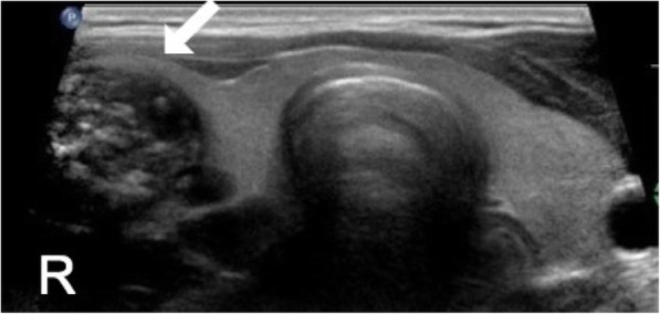
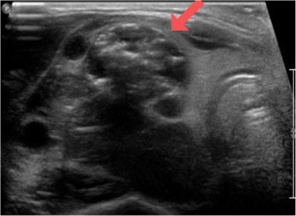
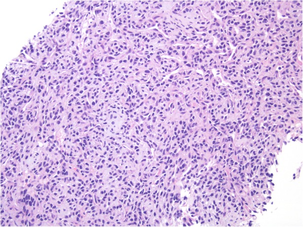
We describe a rare case of mesenchymal chondrosarcoma of the thyroid gland with uncertain primary origin. Thyroid ultrasound showed right sided large, solid, hypoechoic nodule with calcifications and peripheral vascularity and unremarkable isthmus and left thyroid lobe. Fine needle aspiration of the right nodule suggested lymphocytic thyroiditis. The sonographic findings contradicted the typical bilateral clinical and sonographic picture of lymphocytic thyroiditis. A core needle biopsy showed mesenchymal chondrosarcoma.
This case highlights the importance of correlating pathologic diagnosis with sonographic findings, the appropriate utilization of fine needle aspiration and core needle biopsy to evaluate thyroid nodules and the rare incidence of mesenchymal chondrosarcoma involving the thyroid.
甲状腺超声通常用于对偶然发现的甲状腺结节进行风险分层。具有高恶性风险超声特征的结节通过细针穿刺进行评估。粗针活检在甲状腺结节中的作用仅限于细针穿刺结果不明确的病例。
我们描述了一例罕见的甲状腺间叶性软骨肉瘤,原发部位不明。甲状腺超声显示右侧有一个大的实性低回声结节,伴有钙化和周边血管,峡部及左侧甲状腺叶未见明显异常。对右侧结节进行细针穿刺提示淋巴细胞性甲状腺炎。超声检查结果与淋巴细胞性甲状腺炎典型的双侧临床及超声表现不符。粗针活检显示为间叶性软骨肉瘤。
该病例突出了将病理诊断与超声检查结果相关联的重要性,恰当运用细针穿刺和粗针活检来评估甲状腺结节,以及甲状腺发生间叶性软骨肉瘤的罕见性。