• 文献检索
  • 文档翻译
  • 深度研究
  • 学术资讯
  • Suppr Zotero 插件Zotero 插件
  • 邀请有礼
  • 套餐&价格
  • 历史记录
应用&插件
Suppr Zotero 插件Zotero 插件浏览器插件Mac 客户端Windows 客户端微信小程序
定价
高级版会员购买积分包购买API积分包
服务
文献检索文档翻译深度研究API 文档MCP 服务
关于我们
关于 Suppr公司介绍联系我们用户协议隐私条款
关注我们

Suppr 超能文献

核心技术专利:CN118964589B侵权必究
粤ICP备2023148730 号-1Suppr @ 2026

文献检索

告别复杂PubMed语法,用中文像聊天一样搜索,搜遍4000万医学文献。AI智能推荐,让科研检索更轻松。

立即免费搜索

文件翻译

保留排版,准确专业,支持PDF/Word/PPT等文件格式,支持 12+语言互译。

免费翻译文档

深度研究

AI帮你快速写综述,25分钟生成高质量综述,智能提取关键信息,辅助科研写作。

立即免费体验

用于牙髓消毒的工程化生物活性纳米颗粒的抗生物膜和免疫反应

Antibiofilm and Immune Response of Engineered Bioactive Nanoparticles for Endodontic Disinfection.

作者信息

Hussein Hebatullah, Kishen Anil

机构信息

The Kishen Lab, Dental Research Institute, University of Toronto, Toronto, ON M5G 1G6, Canada.

Faculty of Dentistry, University of Toronto, Toronto, ON M5G 1G6, Canada.

出版信息

J Clin Med. 2020 Mar 9;9(3):730. doi: 10.3390/jcm9030730.

DOI:10.3390/jcm9030730
PMID:32182743
原文链接:https://pmc.ncbi.nlm.nih.gov/articles/PMC7141246/
Abstract

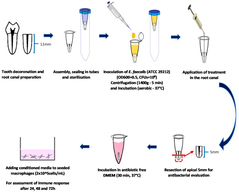
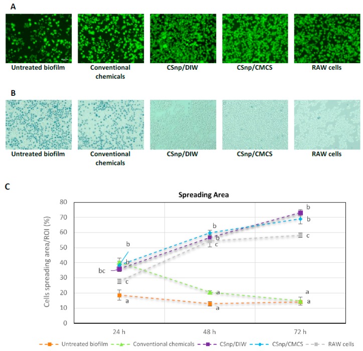
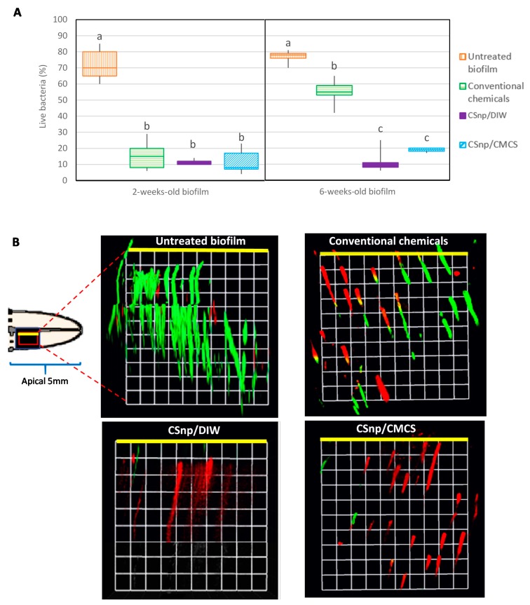
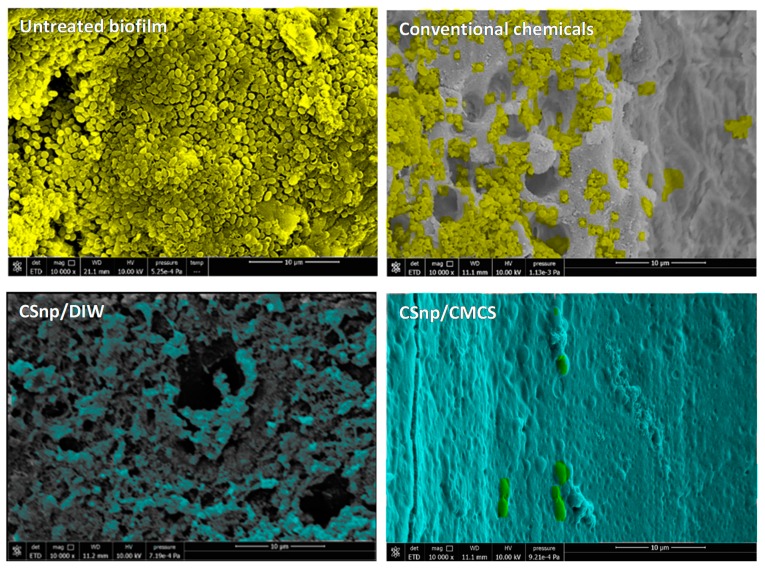
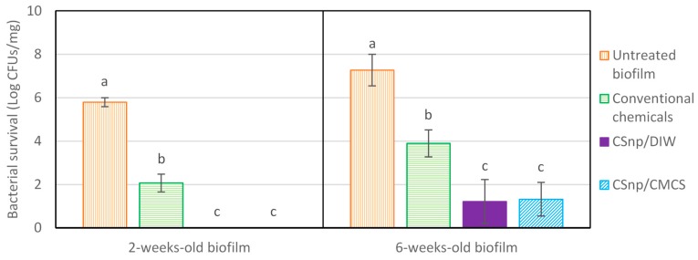
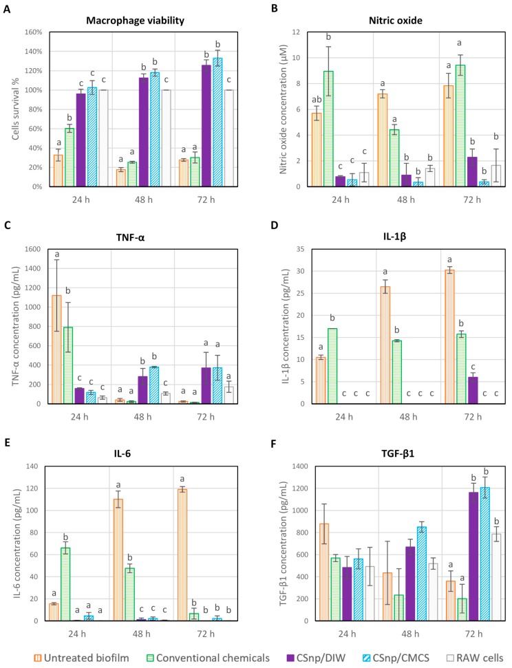
The biological aim of root canal treatment is to facilitate periapical tissue healing following endodontic therapy. This study aimed to develop an organotypic infected root canal model to understand the interaction of bacterial biofilm with macrophages and study the therapeutic effect of engineered bioactive chitosan nanoparticles (CSnp) on macrophages. Ex-vivo experiments were conducted in two phases; Phase-1: biofilms (two and six weeks old) developed in organotypic root canal model were used to characterize residual biofilm after conventional chemical treatment alone and combined with CSnp utilizing Confocal Laser Scanning Microscopy, Scanning Electron Microscopy and colony-forming units from pulverized dentin. Phase-2: The interaction of post-treatment biofilm and RAW macrophages was evaluated regarding pro/anti-inflammatory markers, cell viability and spreading at 24, 48 and 72 h. Compared to conventionally disinfected six-week-old biofilm, CSnp resulted in less viable bacteria ( < 0.01). Scanning electron micrographs demonstrated disruption of the biofilm. CSnp exhibited less residual bacterial load in pulverized dentin ( < 0.001). Macrophage interaction with CSnp-treated biofilm reduced proinflammatory markers (nitric oxide, TNF-α, IL-1β, and IL-6), increased anti-inflammatory marker (TGF-β1) and enhanced cell survival and spreading over time ( < 0.01 at 72 h). Engineered chitosan nanoparticles concurrently inactivated biofilm and altered the inflammatory response of macrophages that would promote healing.

摘要

根管治疗的生物学目的是促进牙髓治疗后根尖周组织的愈合。本研究旨在建立一种器官型感染根管模型,以了解细菌生物膜与巨噬细胞的相互作用,并研究工程化生物活性壳聚糖纳米颗粒(CSnp)对巨噬细胞的治疗效果。体外实验分两个阶段进行;第一阶段:利用共聚焦激光扫描显微镜、扫描电子显微镜和粉碎牙本质中的菌落形成单位,对在器官型根管模型中形成的生物膜(两周和六周龄)进行常规化学处理单独处理以及与CSnp联合处理后,表征残留生物膜。第二阶段:在24、48和72小时评估处理后生物膜与RAW巨噬细胞的相互作用,涉及促炎/抗炎标志物、细胞活力和细胞铺展情况。与传统消毒的六周龄生物膜相比,CSnp导致存活细菌减少(<0.01)。扫描电子显微镜照片显示生物膜受到破坏。CSnp在粉碎牙本质中的残留细菌载量较低(<0.001)。巨噬细胞与经CSnp处理的生物膜相互作用可降低促炎标志物(一氧化氮、TNF-α、IL-1β和IL-6),增加抗炎标志物(TGF-β1),并随着时间的推移提高细胞存活率和促进细胞铺展(72小时时<0.01)。工程化壳聚糖纳米颗粒同时使生物膜失活并改变巨噬细胞的炎症反应,从而促进愈合。

https://cdn.ncbi.nlm.nih.gov/pmc/blobs/75ca/7141246/c4042c26d5de/jcm-09-00730-g006.jpg
https://cdn.ncbi.nlm.nih.gov/pmc/blobs/75ca/7141246/8a9e3de345d9/jcm-09-00730-g001.jpg
https://cdn.ncbi.nlm.nih.gov/pmc/blobs/75ca/7141246/8cb891bbbc80/jcm-09-00730-g002.jpg
https://cdn.ncbi.nlm.nih.gov/pmc/blobs/75ca/7141246/020ce206634e/jcm-09-00730-g003.jpg
https://cdn.ncbi.nlm.nih.gov/pmc/blobs/75ca/7141246/a73f79bf3e08/jcm-09-00730-g004.jpg
https://cdn.ncbi.nlm.nih.gov/pmc/blobs/75ca/7141246/5673c324984f/jcm-09-00730-g005.jpg
https://cdn.ncbi.nlm.nih.gov/pmc/blobs/75ca/7141246/c4042c26d5de/jcm-09-00730-g006.jpg

相似文献

1
Antibiofilm and Immune Response of Engineered Bioactive Nanoparticles for Endodontic Disinfection.用于牙髓消毒的工程化生物活性纳米颗粒的抗生物膜和免疫反应
J Clin Med. 2020 Mar 9;9(3):730. doi: 10.3390/jcm9030730.
2
Engineered Chitosan-based Nanoparticles Modulate Macrophage-Periodontal Ligament Fibroblast Interactions in Biofilm-mediated Inflammation.基于壳聚糖的纳米粒子工程调节生物膜介导的炎症中巨噬细胞-牙周膜成纤维细胞的相互作用。
J Endod. 2021 Sep;47(9):1435-1444. doi: 10.1016/j.joen.2021.06.017. Epub 2021 Jun 29.
3
Proteomic profiling reveals engineered chitosan nanoparticles mediated cellular crosstalk and immunomodulation for therapeutic application in apical periodontitis.蛋白质组学分析揭示了工程化壳聚糖纳米颗粒介导的细胞间串扰和免疫调节在根尖周炎治疗中的应用。
Bioact Mater. 2021 Oct 9;11:77-89. doi: 10.1016/j.bioactmat.2021.09.032. eCollection 2022 May.
4
Antibiofilm efficacy of photosensitizer-functionalized bioactive nanoparticles on multispecies biofilm.光敏剂功能化生物活性纳米颗粒对多物种生物膜的抗生物膜功效
J Endod. 2014 Oct;40(10):1604-10. doi: 10.1016/j.joen.2014.03.009. Epub 2014 Apr 29.
5
Electrokinetic transport and distribution of antibacterial nanoparticles for endodontic disinfection.动电学传输和抗菌纳米颗粒在根管消毒中的分布。
Int Endod J. 2020 Aug;53(8):1120-1130. doi: 10.1111/iej.13321. Epub 2020 Jun 18.
6
Effectiveness of chitosan-propolis nanoparticle against Enterococcus faecalis biofilms in the root canal.壳聚糖-蜂胶纳米粒子对根管内粪肠球菌生物膜的有效性。
BMC Oral Health. 2020 Nov 25;20(1):339. doi: 10.1186/s12903-020-01330-0.
7
Evaluation of the antibacterial efficacy of silver nanoparticles against Enterococcus faecalis biofilm.银纳米颗粒对粪肠球菌生物膜的抗菌效果评估。
J Endod. 2014 Feb;40(2):285-90. doi: 10.1016/j.joen.2013.08.022. Epub 2013 Oct 1.
8
Enhancing antibiofilm activity with functional chitosan nanoparticles targeting biofilm cells and biofilm matrix.用靶向生物膜细胞和生物膜基质的功能性壳聚糖纳米粒子增强抗生物膜活性。
Carbohydr Polym. 2018 Nov 15;200:35-42. doi: 10.1016/j.carbpol.2018.07.072. Epub 2018 Jul 25.
9
Evaluation of Cold Plasma Treatment and Safety in Disinfecting 3-week Root Canal Enterococcus faecalis Biofilm In Vitro.冷等离子体处理对3周龄粪肠球菌根管生物膜体外消毒效果及安全性的评估
J Endod. 2015 Aug;41(8):1325-30. doi: 10.1016/j.joen.2014.10.020. Epub 2015 May 28.
10
Cold plasma therapy of a tooth root canal infected with enterococcus faecalis biofilms in vitro.体外感染粪肠球菌生物膜的根管的冷等离子体治疗。
J Endod. 2013 Jan;39(1):105-10. doi: 10.1016/j.joen.2012.08.017. Epub 2012 Oct 11.

引用本文的文献

1
Advances in hybridized nanoarchitectures for improved oro-dental health.用于改善口腔健康的杂交纳米结构的进展。
J Nanobiotechnology. 2024 Aug 7;22(1):469. doi: 10.1186/s12951-024-02680-5.
2
Exosomes as Promising Therapeutic Tools for Regenerative Endodontic Therapy.外泌体——再生牙髓治疗有前途的治疗工具。
Biomolecules. 2024 Mar 11;14(3):330. doi: 10.3390/biom14030330.
3
Evaluating the Effect of Tideglusib-Loaded Bioactive Glass Nanoparticles as a Potential Dentine Regenerative Material.评估载有替格瑞洛的生物活性玻璃纳米颗粒作为潜在牙本质再生材料的效果。

本文引用的文献

1
Present status and future directions: Microbiology of endodontic infections.现状与未来方向:牙髓感染的微生物学。
Int Endod J. 2022 May;55 Suppl 3:512-530. doi: 10.1111/iej.13677. Epub 2022 Jan 13.
2
Polymeric and inorganic nanoscopical antimicrobial fillers in dentistry.牙科用聚合体和无机纳米抗菌填料。
Acta Biomater. 2020 Jan 1;101:69-101. doi: 10.1016/j.actbio.2019.09.025. Epub 2019 Sep 19.
3
Validation of Biofilm Assays to Assess Antibiofilm Efficacy in Instrumented Root Canals after Syringe Irrigation and Sonic Agitation.
Materials (Basel). 2022 Jun 29;15(13):4567. doi: 10.3390/ma15134567.
4
Antimicrobial and Antibiofilm Properties of Bioceramic Materials in Endodontics.生物陶瓷材料在牙髓病学中的抗菌和抗生物膜特性
Materials (Basel). 2021 Dec 10;14(24):7594. doi: 10.3390/ma14247594.
5
Proteomic profiling reveals engineered chitosan nanoparticles mediated cellular crosstalk and immunomodulation for therapeutic application in apical periodontitis.蛋白质组学分析揭示了工程化壳聚糖纳米颗粒介导的细胞间串扰和免疫调节在根尖周炎治疗中的应用。
Bioact Mater. 2021 Oct 9;11:77-89. doi: 10.1016/j.bioactmat.2021.09.032. eCollection 2022 May.
6
Nonsurgical endodontic management of mandibular first premolar with incomplete root-end resection.下颌第一前磨牙牙根尖切除不完整的非手术牙髓治疗管理
Clin Case Rep. 2020 Nov 29;9(1):437-444. doi: 10.1002/ccr3.3551. eCollection 2021 Jan.
7
New Ways to Disinfect and Fill Dental Hard Tissues.牙科硬组织消毒与填充的新方法
J Clin Med. 2020 May 18;9(5):1524. doi: 10.3390/jcm9051524.
经注射器冲洗和超声震荡后评估器械根管内抗生物膜功效的生物膜检测方法的验证。
J Endod. 2018 Feb;44(2):292-298. doi: 10.1016/j.joen.2017.10.005. Epub 2017 Dec 16.
4
Intracellular disposition of chitosan nanoparticles in macrophages: intracellular uptake, exocytosis, and intercellular transport.壳聚糖纳米颗粒在巨噬细胞中的细胞内分布:细胞内摄取、胞吐作用和细胞间转运。
Int J Nanomedicine. 2017 Aug 31;12:6383-6398. doi: 10.2147/IJN.S142060. eCollection 2017.
5
Effects of a Bioactive Scaffold Containing a Sustained Transforming Growth Factor-β1-releasing Nanoparticle System on the Migration and Differentiation of Stem Cells from the Apical Papilla.一种含有持续释放转化生长因子-β1纳米颗粒系统的生物活性支架对根尖乳头干细胞迁移和分化的影响。
J Endod. 2016 Sep;42(9):1385-92. doi: 10.1016/j.joen.2016.06.017. Epub 2016 Jul 30.
6
Characterizing the collagen stabilizing effect of crosslinked chitosan nanoparticles against collagenase degradation.表征交联壳聚糖纳米颗粒对胶原酶降解的胶原稳定作用。
Dent Mater. 2016 Aug;32(8):968-77. doi: 10.1016/j.dental.2016.05.005. Epub 2016 May 31.
7
Surface modification of nanoparticles enables selective evasion of phagocytic clearance by distinct macrophage phenotypes.纳米颗粒的表面修饰使它们能够有选择地逃避不同巨噬细胞表型的吞噬清除。
Sci Rep. 2016 May 19;6:26269. doi: 10.1038/srep26269.
8
Nanoparticles for antimicrobial purposes in Endodontics: A systematic review of in vitro studies.用于牙髓病学抗菌目的的纳米颗粒:体外研究的系统评价
Mater Sci Eng C Mater Biol Appl. 2016 Jan 1;58:1269-78. doi: 10.1016/j.msec.2015.08.070. Epub 2015 Sep 4.
9
Correlation between Enterococcus faecalis and Persistent Intraradicular Infection Compared with Primary Intraradicular Infection: A Systematic Review.粪肠球菌与根管内持续感染的相关性与原发性根管内感染的比较:一项系统评价
J Endod. 2015 Aug;41(8):1207-13. doi: 10.1016/j.joen.2015.04.008. Epub 2015 May 23.
10
Temporal-controlled Dexamethasone Releasing Chitosan Nanoparticle System Enhances Odontogenic Differentiation of Stem Cells from Apical Papilla.时间控制的地塞米松释放壳聚糖纳米颗粒系统增强根尖乳头干细胞的成牙分化
J Endod. 2015 Aug;41(8):1253-8. doi: 10.1016/j.joen.2015.03.024. Epub 2015 May 5.