Ophthalmology Group, IRIBHM (Institut de Recherche Interdisciplinaire en Biologie Humaine et Moléculaire), Université Libre de Bruxelles (ULB), Erasme Campus, Building C, Room C6.117, 808 Route de Lennik, 1070, Brussels, Belgium.
Ophthalmology Department of Erasme Hospital, Université Libre de Bruxelles (ULB), 808 Route de Lennik, 1070, Brussels, Belgium.
BMC Ophthalmol. 2020 Mar 17;20(1):106. doi: 10.1186/s12886-020-1333-5.
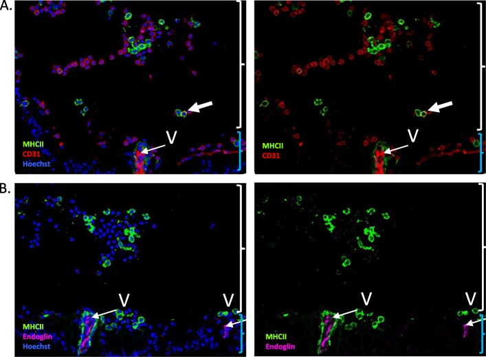
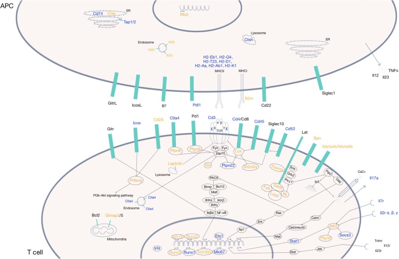
Blood-retinal barrier cells are known to exhibit a massive phenotypic change during experimental autoimmune uveitis (EAU) development. In an attempt to investigate the mechanisms of blood-retinal barrier (BRB) breakdown at a global level, we studied the gene regulation of total retinal cells and retinal endothelial cells during non-infectious uveitis.
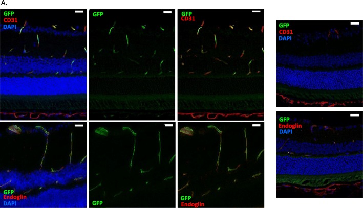
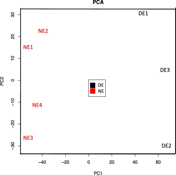
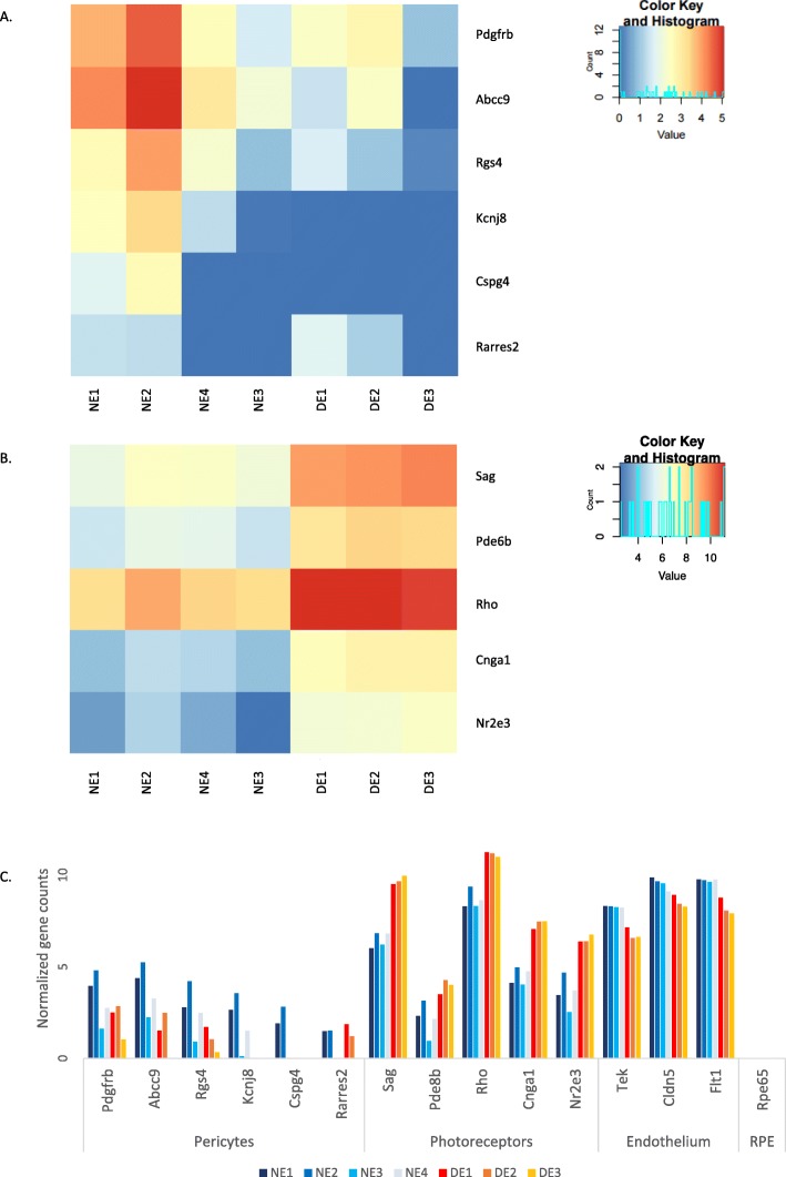
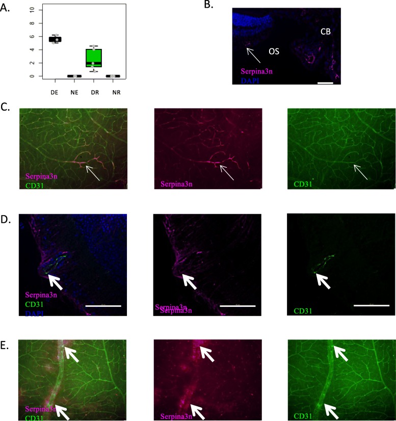
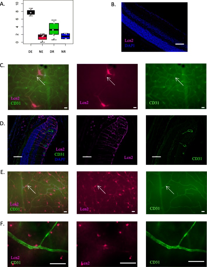
Retinal endothelial cells were isolated by flow cytometry either in Tie2-GFP mice (CD31 CD45 GFP cells), or in wild type C57BL/6 mice (CD31 CD45 endoglin cells). EAU was induced in C57BL/6 mice by adoptive transfer of IRBP1-20-specific T cells. Total retinal cells and retinal endothelial cells from naïve and EAU mice were sorted and their gene expression compared by RNA-Seq. Protein expression of selected genes was validated by immunofluorescence on retinal wholemounts and cryosections and by flow cytometry.
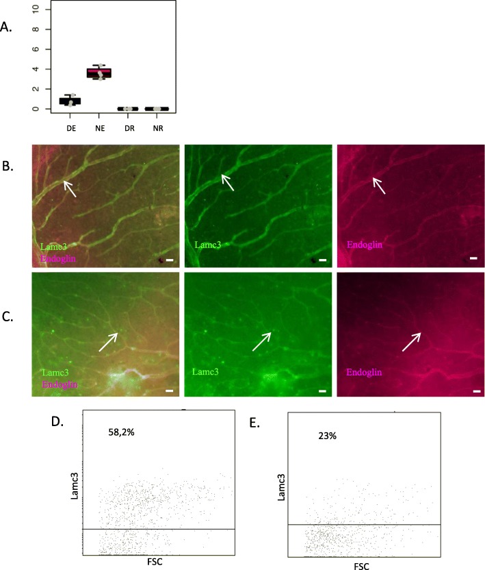
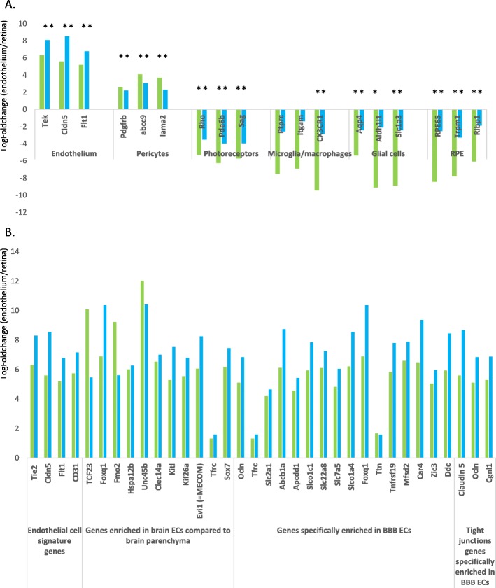
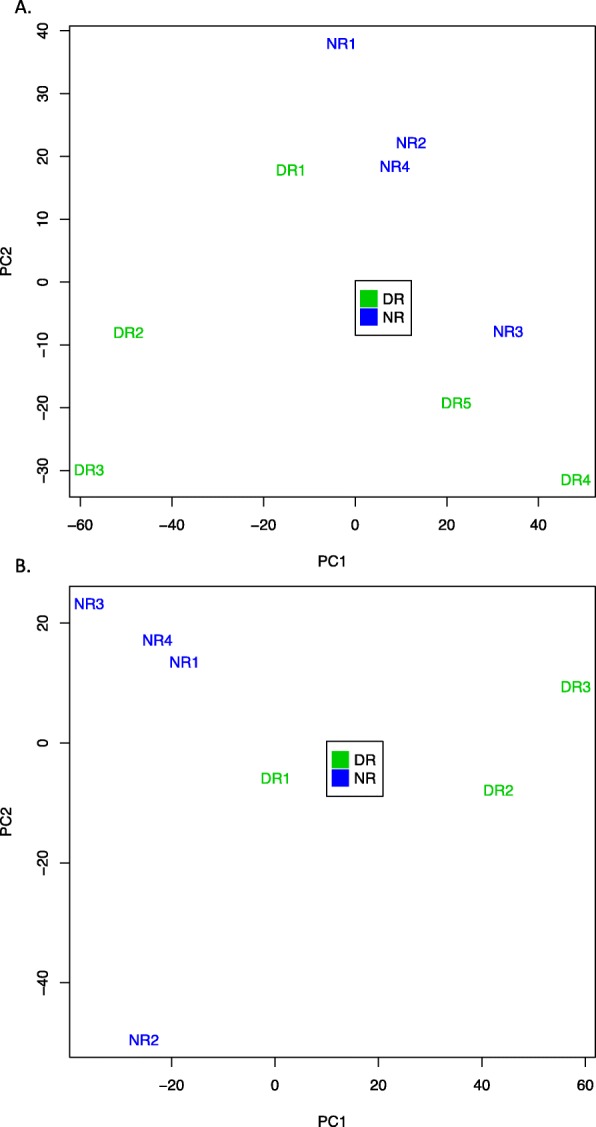
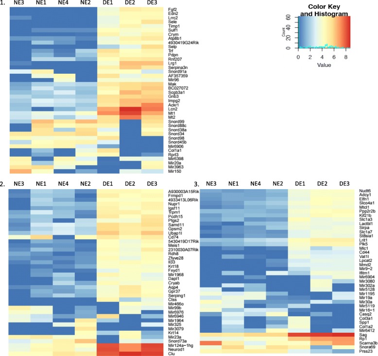
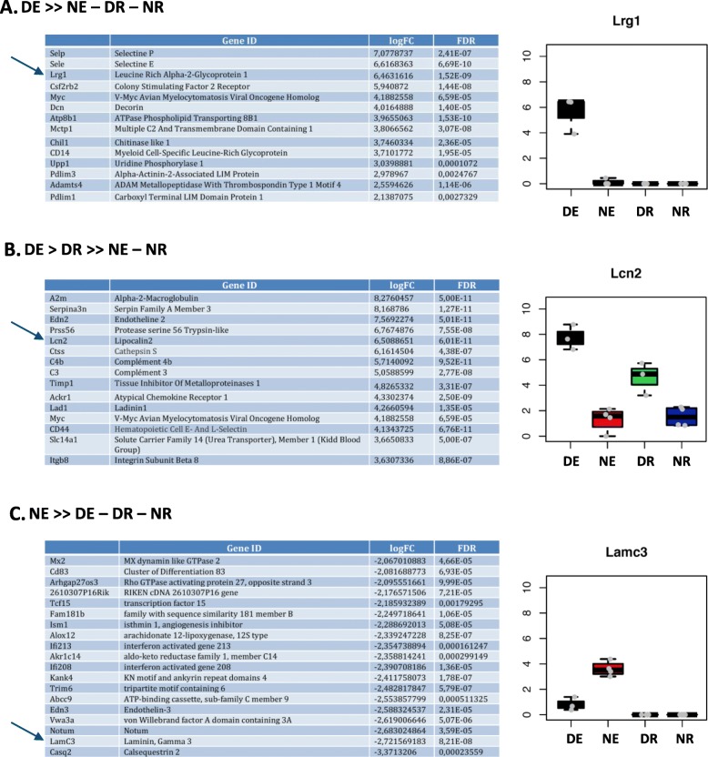
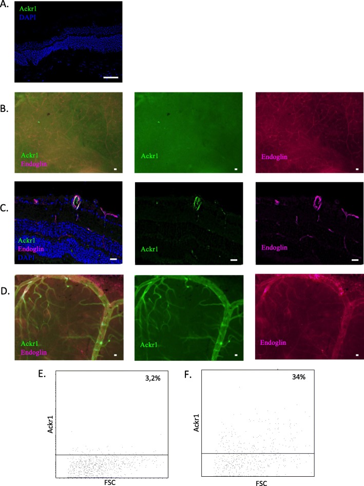
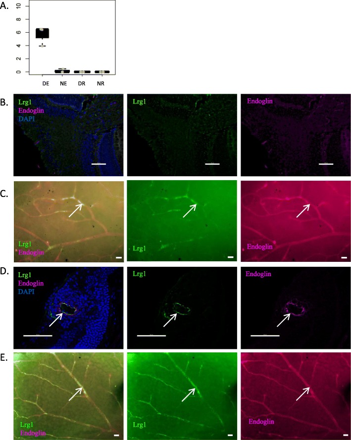
Retinal endothelial cell sorting in wild type C57BL/6 mice was validated by comparative transcriptome analysis with retinal endothelial cells sorted from Tie2-GFP mice, which express GFP under the control of the endothelial-specific receptor tyrosine kinase promoter Tie2. RNA-Seq analysis of total retinal cells mainly brought to light upregulation of genes involved in antigen presentation and T cell activation during EAU. Specific transcriptome analysis of retinal endothelial cells allowed us to identify 82 genes modulated in retinal endothelial cells during EAU development. Protein expression of 5 of those genes (serpina3n, lcn2, ackr1, lrg1 and lamc3) was validated at the level of inner BRB cells.
Those data not only confirm the involvement of known pathogenic molecules but further provide a list of new candidate genes and pathways possibly implicated in inner BRB breakdown during non-infectious posterior uveitis.
已知血视网膜屏障细胞在实验性自身免疫性葡萄膜炎 (EAU) 发展过程中会发生大量表型变化。为了从全局水平研究血视网膜屏障 (BRB) 破坏的机制,我们研究了非传染性葡萄膜炎期间整个视网膜细胞和视网膜内皮细胞的基因调控。
通过流式细胞术从 Tie2-GFP 小鼠(CD31 CD45 GFP 细胞)或野生型 C57BL/6 小鼠(CD31 CD45 endoglin 细胞)中分离视网膜内皮细胞。通过过继转移 IRBP1-20 特异性 T 细胞在 C57BL/6 小鼠中诱导 EAU。从 naive 和 EAU 小鼠中分离出总视网膜细胞和视网膜内皮细胞,并通过 RNA-Seq 比较它们的基因表达。通过视网膜全层和冷冻切片免疫荧光和流式细胞术验证选定基因的蛋白表达。
通过与从 Tie2-GFP 小鼠中分离的视网膜内皮细胞进行比较转录组分析,验证了野生型 C57BL/6 小鼠中视网膜内皮细胞的分选,该细胞在内皮特异性受体酪氨酸激酶启动子 Tie2 的控制下表达 GFP。EAU 期间,总视网膜细胞的 RNA-Seq 分析主要揭示了参与抗原呈递和 T 细胞激活的基因上调。视网膜内皮细胞的特异性转录组分析使我们能够鉴定出 82 个在 EAU 发展过程中在视网膜内皮细胞中被调节的基因。其中 5 个基因(serpina3n、lcn2、ackr1、lrg1 和 lamc3)的蛋白表达在内 BRB 细胞水平上得到验证。
这些数据不仅证实了已知致病分子的参与,而且进一步提供了一系列可能参与非传染性后葡萄膜炎中内 BRB 破坏的新候选基因和途径。