Thiriot Aude, Perdomo Carolina, Cheng Guiying, Novitzky-Basso Igor, McArdle Sara, Kishimoto Jamie K, Barreiro Olga, Mazo Irina, Triboulet Robinson, Ley Klaus, Rot Antal, von Andrian Ulrich H
Department of Microbiology and Immunobiology & HMS Center for Immune Imaging, Harvard Medical School, 77 Avenue Louis Pasteur, Boston, MA, 02115, USA.
The Ragon Institute of MGH, MIT and Harvard, Cambridge, MA, 02139, USA.
BMC Biol. 2017 May 19;15(1):45. doi: 10.1186/s12915-017-0381-7.
Intravascular leukocyte recruitment in most vertebrate tissues is restricted to postcapillary and collecting venules, whereas capillaries and arterioles usually support little or no leukocyte adhesion. This segmental restriction is thought to be mediated by endothelial, rather than hemodynamic, differences. The underlying mechanisms are largely unknown, in part because effective tools to distinguish, isolate, and analyze venular endothelial cells (V-ECs) and non-venular endothelial cells (NV-ECs) have been unavailable. We hypothesized that the atypical chemokine receptor DARC (Duffy Antigen Receptor for Chemokines, a.k.a. ACKR1 or CD234) may distinguish V-ECs versus NV-ECs in mice.
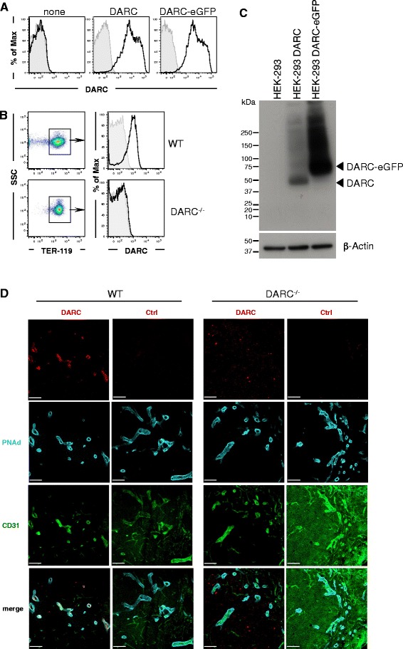
We generated a rat-anti-mouse monoclonal antibody (MAb) that specifically recognizes the erythroid and endothelial forms of native, surface-expressed DARC. Using this reagent, we characterized DARC expression and distribution in the microvasculature of murine tissues.
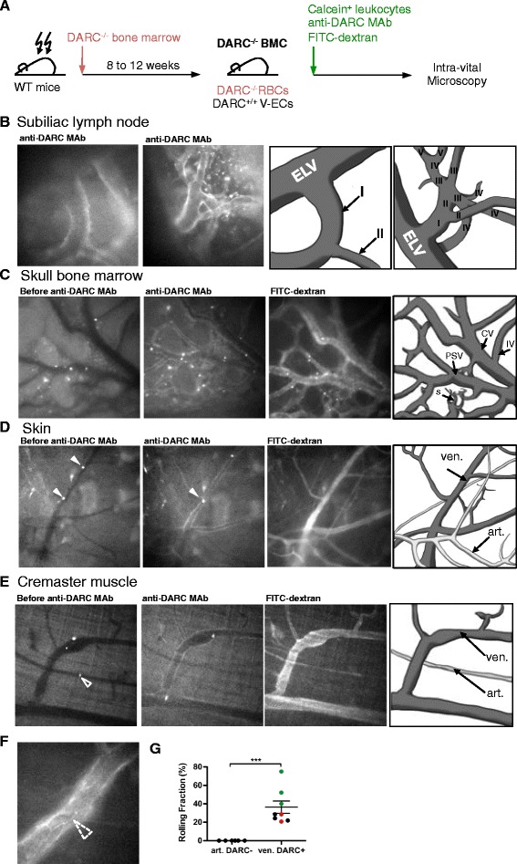
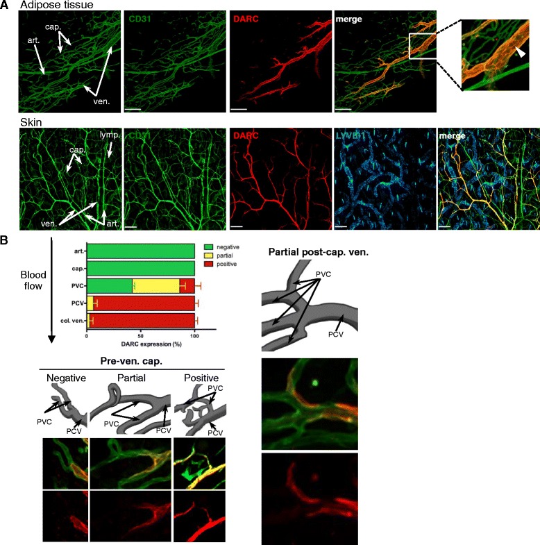
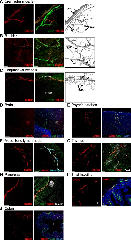
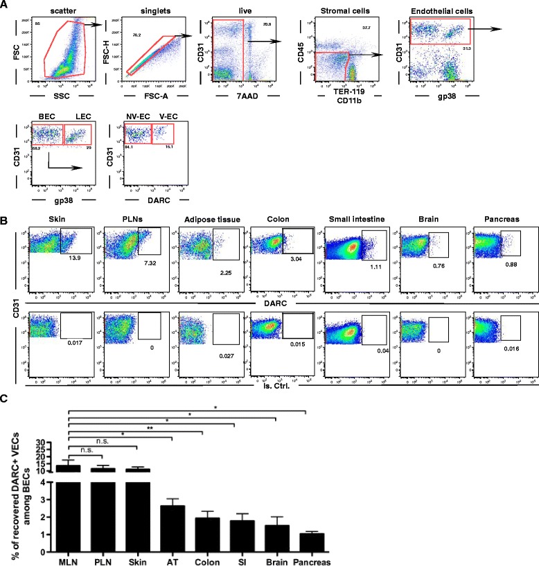
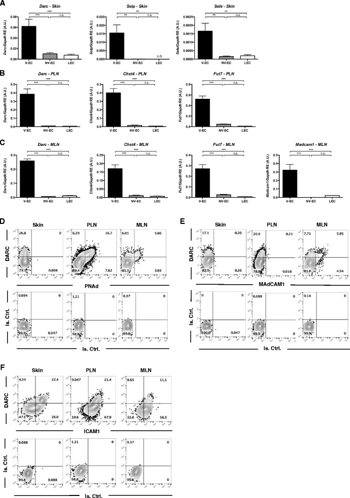
DARC was exquisitely restricted to post-capillary and small collecting venules and completely absent from arteries, arterioles, capillaries, veins, and most lymphatics in every tissue analyzed. Accordingly, intravital microscopy showed that adhesive leukocyte-endothelial interactions were restricted to DARC venules. DARC was detectable over the entire circumference of V-ECs, but was more concentrated at cell-cell junctions. Analysis of single-cell suspensions suggested that the frequency of V-ECs among the total microvascular EC pool varies considerably between different tissues.
Immunostaining of endothelial DARC allows the identification and isolation of intact V-ECs from multiple murine tissues. This strategy may be useful to dissect the mechanisms underlying segmental microvascular specialization in healthy and diseased tissues and to characterize the role of EC subsets in tissue-homeostasis, immune surveillance, infection, inflammation, and malignancies.
在大多数脊椎动物组织中,血管内白细胞募集仅限于毛细血管后微静脉和集合微静脉,而毛细血管和小动脉通常很少或几乎没有白细胞黏附。这种节段性限制被认为是由内皮细胞差异介导的,而非血流动力学差异。其潜在机制在很大程度上尚不清楚,部分原因是缺乏有效工具来区分、分离和分析微静脉内皮细胞(V-ECs)和非微静脉内皮细胞(NV-ECs)。我们推测非典型趋化因子受体DARC(趋化因子的达菲抗原受体,又名ACKR1或CD234)可能区分小鼠的V-ECs和NV-ECs。
我们制备了一种大鼠抗小鼠单克隆抗体(MAb),该抗体能特异性识别天然的、表面表达的DARC的红细胞和内皮细胞形式。使用该试剂,我们对DARC在小鼠组织微血管中的表达和分布进行了表征。
在每个分析的组织中,DARC精确地局限于毛细血管后微静脉和小集合微静脉,在动脉、小动脉、毛细血管、静脉和大多数淋巴管中完全不存在。因此,活体显微镜检查显示黏附性白细胞-内皮细胞相互作用局限于表达DARC的微静脉。DARC在V-ECs的整个圆周上都可检测到,但在细胞间连接处更为集中。单细胞悬液分析表明,在不同组织中,V-ECs在总微血管内皮细胞池中的频率差异很大。
内皮DARC的免疫染色可从多种小鼠组织中识别和分离完整的V-ECs。该策略可能有助于剖析健康和患病组织中节段性微血管特化的潜在机制,并表征内皮细胞亚群在组织稳态、免疫监视、感染、炎症和恶性肿瘤中的作用。