Gozzo Cecilia, Giambelluca Dario, Cannella Roberto, Caruana Giovanni, Jukna Agita, Picone Dario, Midiri Massimo, Salvaggio Giuseppe
Sezione di Scienze Radiologiche, Biomedicina, Neuroscienze e Diagnostica avanzata (BIND), University of Palermo, Via del Vespro 129, 90127, Palermo, Italy.
Department of Medical Surgical Sciences and Advanced Technologies "G.F. Ingrassia"-Radiology I Unit, University Hospital "Policlinico Vittorio Emanuele", Catania, Italy.
Insights Imaging. 2020 Mar 17;11(1):48. doi: 10.1186/s13244-020-00852-z.
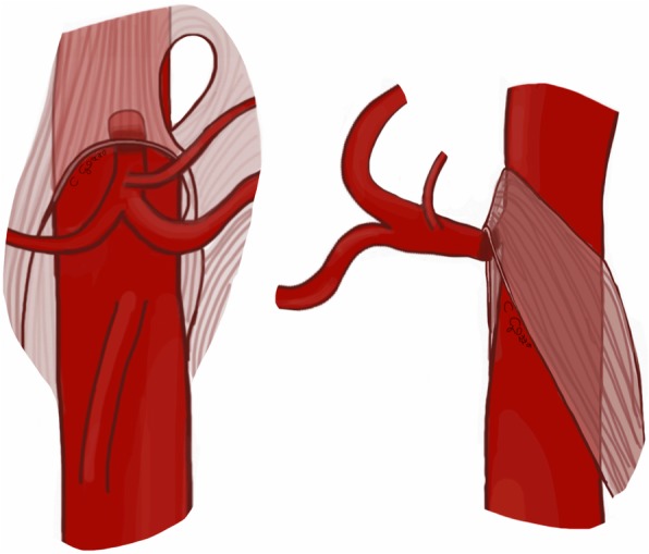
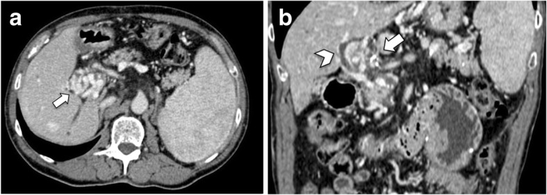
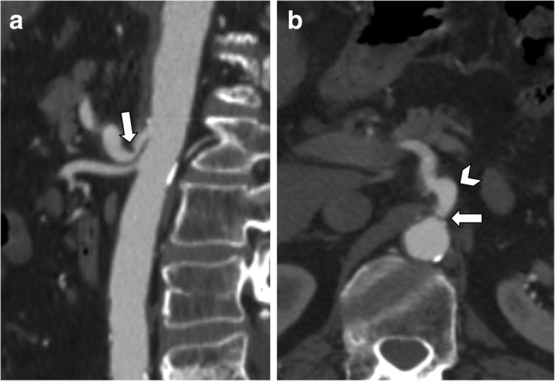
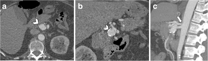
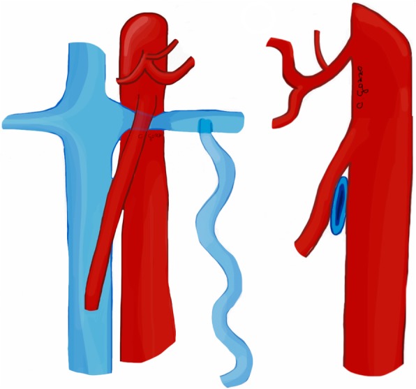
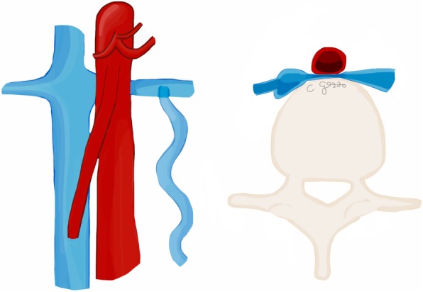
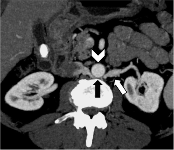
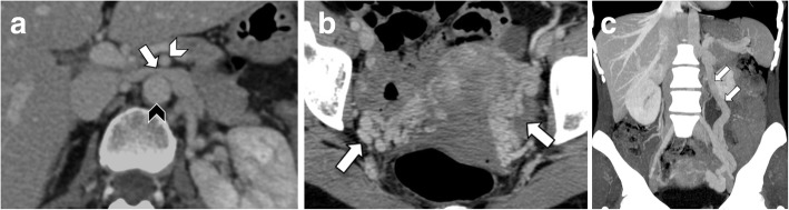
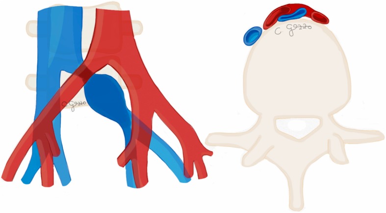
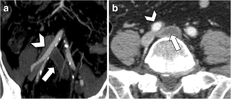
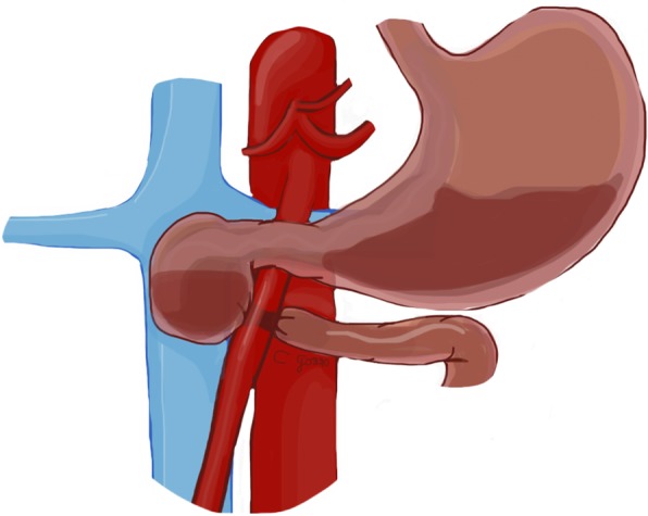
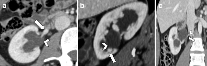
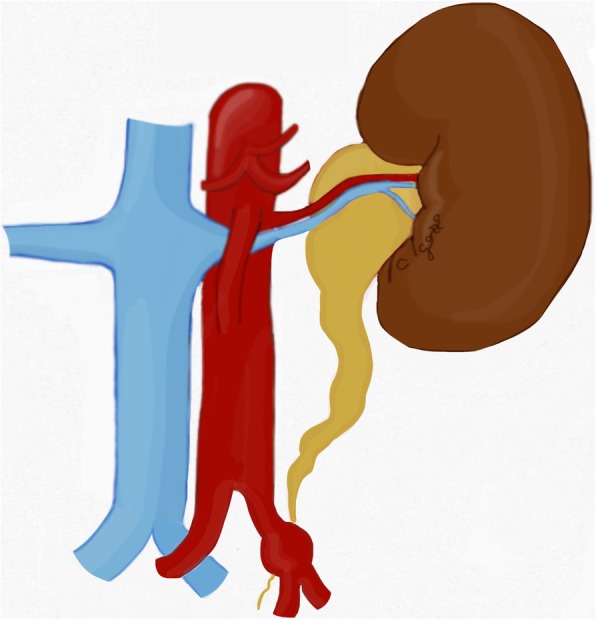
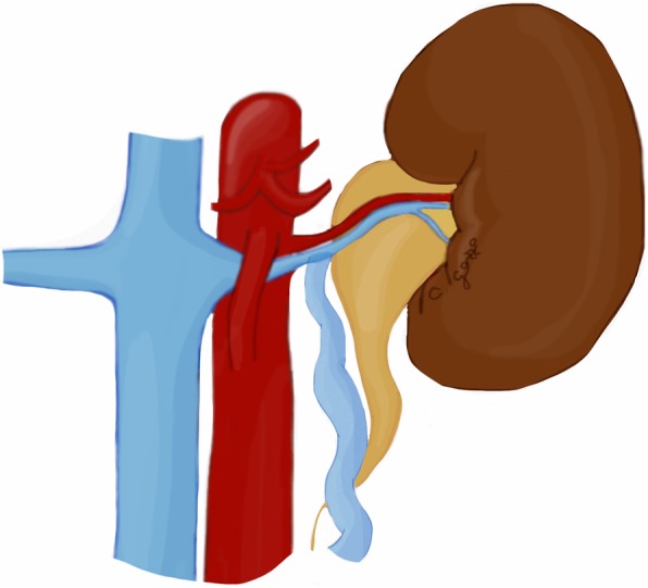
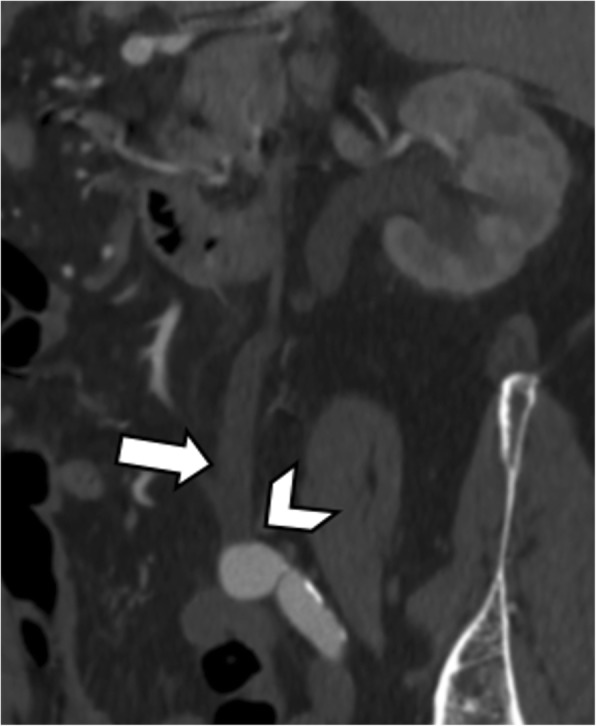
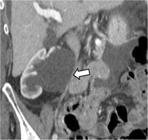
Abdominopelvic vascular compression syndromes include a variety of uncommon conditions characterized by either extrinsic compression of blood vessels by adjacent anatomical structures (i.e., median arcuate ligament syndrome, nutcracker syndrome, May-Thurner syndrome) or compression of hollow viscera by adjacent vessels (i.e., superior mesenteric artery syndrome, ureteropelvic junction obstruction, ureteral vascular compression syndromes, portal biliopathy). These syndromes can be unexpectedly diagnosed even in asymptomatic patients and the predisposing anatomic conditions can be incidentally discovered on imaging examinations performed for other indications, or they can manifest with atypical abdominal symptoms and acute complications, which may lead to significant morbidity if unrecognized. Although computed tomography (CT) is an accurate noninvasive technique for their detection, the diagnosis remains challenging due to the uncommon clinical presentation and often overlooked imaging features. Dynamic imaging may be performed in order to evaluate patients with inconstant symptoms manifesting in a specific position. The purposes of this paper are to review the CT imaging findings of abdominopelvic vascular compression syndromes, correlating with anatomical variants and to provide key features for the noninvasive imaging diagnosis.
腹盆腔血管压迫综合征包括多种罕见病症,其特征为相邻解剖结构对外周血管的外在压迫(即正中弓状韧带综合征、胡桃夹综合征、梅-图二氏综合征)或相邻血管对中空脏器的压迫(即肠系膜上动脉综合征、肾盂输尿管连接部梗阻、输尿管血管压迫综合征、门静脉性肝病)。即使在无症状患者中也可能意外诊断出这些综合征,其易感解剖状况可能在因其他指征进行的影像学检查中偶然发现,或者它们可能表现为非典型腹部症状和急性并发症,如果未被识别,可能导致严重的发病率。尽管计算机断层扫描(CT)是检测这些综合征的准确无创技术,但由于临床表现罕见且影像学特征常被忽视,诊断仍然具有挑战性。对于在特定体位出现不恒定症状的患者,可能需要进行动态成像检查。本文的目的是回顾腹盆腔血管压迫综合征的CT影像学表现,与解剖变异相关联,并为无创影像学诊断提供关键特征。