Department of Orthopaedic Surgery, Children's Hospital of Nanjing Medical University, Nanjing, 210000, Jiangsu Province, China.
J Orthop Surg Res. 2020 Mar 19;15(1):111. doi: 10.1186/s13018-020-01615-8.
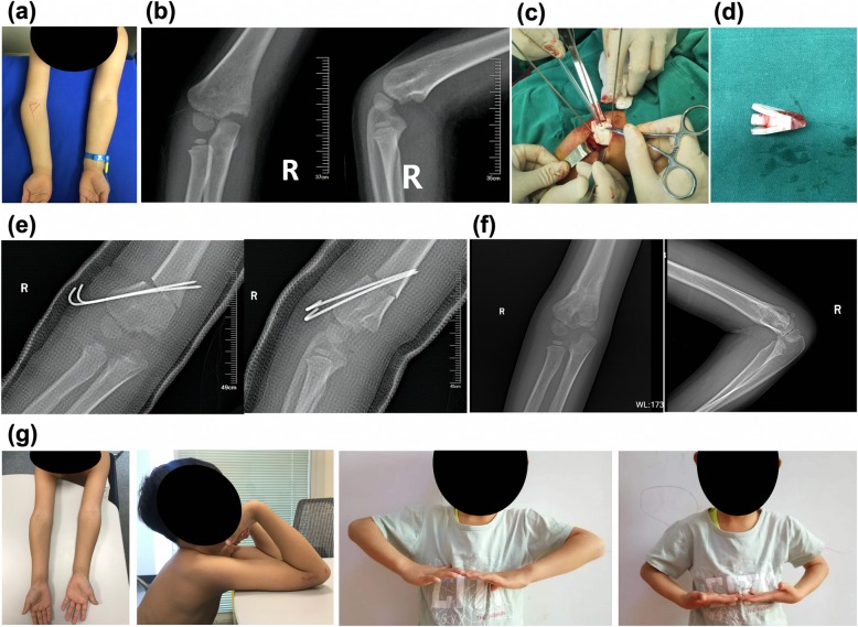
Cubitus varus deformity is a common sequela of elbow fractures in children. Cubitus varus deformity treatment is tending toward 3D correction, which is challenging for orthopedic surgeons. This study aims to explore whether individualized 3D-printed navigation templates can assist with accurate and effective corrective treatment of children with cubitus varus deformity.
Thirty-five patients were treated for cubitus varus deformity from June 2015 to April 2017, including 21 boys and 14 girls, aged 4.6-13.2 years (average, 7.5 years). Of these cases, 17 deformities were on the left side and 18 were on the right side. All were treated with wedge osteotomy of the lateral distal humerus. 3D-printed navigation templates were used in 16 cases, while traditional surgery was used in 19 cases. All patients underwent computed tomography scans before surgery. Computer software was used to analyze the measurements and design and print individualized navigation templates. The navigation templates were matched, and surgery was initially simulated. Intraoperative individualized navigation templates were used to assist with accurate osteotomy and Kirschner wire fixation. Operation times were recorded in all cases, the carrying angles before and after surgery were assessed by computer, and postoperative elbow joint function was evaluated using Bellemore criteria. All measurement data were presented as means ± SD, and Student's t test was used to examine differences between groups. All count data between both groups were compared using the chi-square test or Fisher's exact test analysis.
All individualized navigation templates matched well with the corresponding anatomical markers and were consistent with preoperative planning, simulated surgery, and intraoperative procedures. Average operation times from clear exposure to fixed Kirschner wire were 11.69 min (9.6-13.5 min) for the individualized navigation template group and 22.89 min (17.7-26.8 min) for the traditional operation group (p < 0.001). Average differences in postoperation carrying angles between affected and healthy sides were 1.13° (0-2.0°) and 4.21° (0-7.5°), respectively (p < 0.001). Follow-up 6-12 months postoperation showed that elbow function did not differ significantly between groups using the Bellemore criteria (p > 0.05).
Individualized navigation templates simplify procedures, reduce operation time, and improve accuracy when used in orthopedic surgery to treat children with cubitus varus deformity.
肘内翻畸形是儿童肘部骨折的常见后遗症。肘内翻畸形的治疗倾向于 3D 矫正,这对骨科医生来说具有挑战性。本研究旨在探讨个体化 3D 打印导航模板是否有助于准确有效地矫正儿童肘内翻畸形。
2015 年 6 月至 2017 年 4 月,共收治 35 例儿童肘内翻畸形患者,其中男 21 例,女 14 例;年龄 4.6-13.2 岁,平均 7.5 岁。左侧 17 例,右侧 18 例。所有患者均行肱骨外髁楔形截骨术。其中 16 例行 3D 打印导航模板手术,19 例行传统手术。所有患者术前均行 CT 扫描。使用计算机软件分析测量值并设计和打印个体化导航模板。模板匹配,初步模拟手术。术中使用个体化导航模板辅助精确截骨和克氏针固定。记录所有患者的手术时间,用计算机评估术前、术后提携角,并采用 Bellemore 标准评价术后肘关节功能。所有测量数据均以均数±标准差表示,组间比较采用 Student's t 检验。组间计数资料比较采用卡方检验或 Fisher 确切概率法。
所有个体化导航模板均与相应解剖标志匹配良好,与术前规划、模拟手术和术中操作一致。个体化导航模板组从显露至固定克氏针的平均手术时间为 11.69 分钟(9.6-13.5 分钟),传统手术组为 22.89 分钟(17.7-26.8 分钟)(p<0.001)。术后患侧与健侧携带角的平均差值分别为 1.13°(0-2.0°)和 4.21°(0-7.5°)(p<0.001)。术后 6-12 个月随访时,采用 Bellemore 标准评价两组肘关节功能无显著差异(p>0.05)。
在治疗儿童肘内翻畸形的骨科手术中,个体化导航模板简化了手术步骤,缩短了手术时间,提高了手术准确性。