Silva-Adaya Daniela, Ramos-Chávez Lucio Antonio, Petrosyan Pavel, González-Alfonso Wendy Leslie, Pérez-Acosta Alegna, Gonsebatt Maria E
Departamento de Medicina Genómica, Instituto de Investigaciones Biomédicas, Universidad Nacional Autónoma de México, México, Mexico.
Laboratorio Experimental de Enfermedades Neurodegenerativas, Instituto Nacional de Neurología y Neurocirugía, México, Mexico.
Front Cell Neurosci. 2020 Feb 25;14:17. doi: 10.3389/fncel.2020.00017. eCollection 2020.
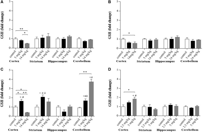
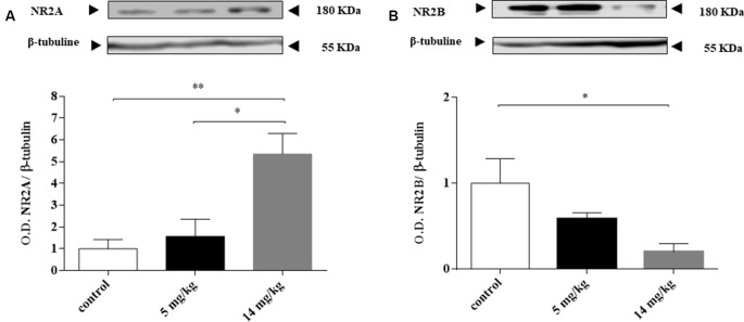
Exposure to toxic metals and metalloids is an important cause of preventable diseases worldwide. Inorganic arsenic (iAs) affects several organs and tissues, causing neurobehavioral alterations in the central nervous system (CNS) that might lead to neurodegeneration. In this work, we wanted to explore the time- and dose-related changes on glutathione (GSH) levels in several regions of the CNS, such as the cortex, striatum, hippocampus, and cerebellum, to identify the initial cellular changes associated to GSH depletion due to iAs exposure. Mice received a single intraperitoneal injection containing 5 or 14 mg/kg sodium arsenite. Animals were killed at 2, 6, and 24 h. Significant depletion of GSH levels was observed in the cortex at 2 and 6 h, while on the striatum, hippocampus, or cerebellum regions, no significant changes were observed. GSH depletion in the cortex was associated with the activation of the nuclear factor erythroid 2-related factor 2 (Nrf2) and nuclear factor kappa B (NFκB) pathways, which led to the upregulation of xCT, excitatory amino acid carrier 1 (EAAC1), glutamate/aspartate transporter (GLAST), and glial glutamate transporter 1 (GLT-1), and the activation of the transsulfuration pathways, which led to the overproduction of HS in the cortex and increased levels of GSH in the cortex and cerebellum at 24 h. In the cortex, the -methyl-D-aspartate (NMDA) receptor subunits NR2A and NR2B were also altered at 24 h. These early effects were not homogeneous among different brain regions and indicate early neurotoxic alterations in the cortex and cerebellum.
接触有毒金属和类金属是全球可预防疾病的一个重要原因。无机砷(iAs)会影响多个器官和组织,导致中枢神经系统(CNS)出现神经行为改变,这可能会引发神经退行性变。在这项研究中,我们想要探究CNS多个区域(如皮层、纹状体、海马体和小脑)中谷胱甘肽(GSH)水平随时间和剂量的变化,以确定与iAs暴露导致的GSH耗竭相关的初始细胞变化。小鼠接受一次腹腔注射,注射物含5或14mg/kg的亚砷酸钠。在2、6和24小时处死动物。在2小时和6小时时,皮层中观察到GSH水平显著降低,而在纹状体、海马体或小脑区域未观察到显著变化。皮层中GSH的耗竭与核因子红细胞2相关因子2(Nrf2)和核因子κB(NFκB)信号通路的激活有关,这导致xCT、兴奋性氨基酸转运体1(EAAC1)、谷氨酸/天冬氨酸转运体(GLAST)和胶质谷氨酸转运体1(GLT-1)上调,以及转硫途径的激活,这导致24小时时皮层中HS过量产生,皮层和小脑中GSH水平升高。在皮层中,24小时时N-甲基-D-天冬氨酸(NMDA)受体亚基NR2A和NR2B也发生了改变。这些早期效应在不同脑区并不一致,表明皮层和小脑出现了早期神经毒性改变。