De Alok, De Archana, Sharma Ramratan, Suo William, Sharma Mukut
Kansas City VA Medical Center and Midwest Veterans Biomedical Research Foundation, Kansas City, MO 64128, USA.
J Cancer. 2020 Jan 29;11(7):1927-1939. doi: 10.7150/jca.36919. eCollection 2020.
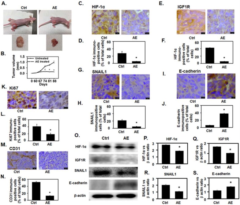
Ovarian cancer (OC), the most lethal gynecologic malignancy, is highly resistant to current treatment strategies. High-grade serous epithelial ovarian cancer (HGSOC) cells with increased somatic mutations and genomic instability and the resulting heterogeneous mutant phenotypes are highly resistant to therapy. Plant-derived natural products, including Amla () extract (AE), have demonstrated potent anti-neoplastic properties. Recently we demonstrated that AE inhibits cell growth and the expression of angiogenic factors in OVCAR3 and SKOV3 OC cells as well as in xenografts . The goal of this study was to determine the anti-proliferative, anti-angiogenic and anti-metastatic effects of AE on carboplatinum- and taxol-resistant HGSOC cells carrying p53, BRCA1/2 mutations. Anti-proliferative and anti-metastatic effects of AE on recently characterized carboplatinum- and taxol-resistant HGSOC cells (TOV3041G, OV866(2), OV4453 and, OV4485) was determined using the MTT, migration, invasion and spheroid assays i To understand the mechanism of AE-induced changes in angiogenesis-related hypoxia-inducible factor 1α (HIF-1α) and insulin growth factor receptor 1 (IGF1R), and EMT-associated SNAIL1 and E-cadherin proteins were studied using immunostaining and Western blotting. effects of AE were determined using mouse xenograft tumor model of OC developed by subcutaneous injection of OV4485 cells that carry mutant p53 and BRCA1, most aggressive and resistant among HGSOC cell lines used in this study. Tumor growth was measured using morphometry. Immunostaining and Western blotting were used to determine changes in Ki67 (proliferation marker), CD31 (angiogenesis marker) as well as changes in HIF-1α, IGF1R, SNAIL1 and E-cadherin proteins. AE significantly attenuated migration and invasiveness properties of all tested HGSOC cell phenotypes (P≤0.001), significantly reduced the expression of HIF-1α, IGF1R, and SNAIL1 and increased the expression of E-cadherin in all tested HGSOC cell lines (P=<0.05). Oral administration of AE for 4 weeks caused a significant regression of mouse xenograft tumor (>60%) that derived from OV4855 cells and decreased the expression of endothelial cell antigen-CD31, HIF-1α, IGF1R and SNAIL1 and increased the expression of E-cadherin in tumor tissues. AE sensitizes platinum- and taxol-resistant heterogenous HGSOC cells carrying mutations in p53, BRCA1/2 genes, and attenuates their malignant characteristics through targeting key signaling mechanisms of angiogenesis and metastasis. AE is a potential adjunct therapeutic agent for treating resistant, mutant, heterogenous OC.
卵巢癌(OC)是最致命的妇科恶性肿瘤,对当前的治疗策略具有高度抗性。具有体细胞突变增加和基因组不稳定以及由此产生的异质突变表型的高级别浆液性上皮性卵巢癌(HGSOC)细胞对治疗具有高度抗性。包括余甘子提取物(AE)在内的植物源天然产物已显示出强大的抗肿瘤特性。最近我们证明,AE可抑制OVCAR3和SKOV3 OC细胞以及异种移植瘤中细胞的生长和血管生成因子的表达。本研究的目的是确定AE对携带p53、BRCA1/2突变的卡铂和紫杉醇耐药HGSOC细胞的抗增殖、抗血管生成和抗转移作用。使用MTT、迁移、侵袭和球体测定法确定AE对最近鉴定的卡铂和紫杉醇耐药HGSOC细胞(TOV3041G、OV866(2)、OV4453和OV4485)的抗增殖和抗转移作用。为了解AE诱导的血管生成相关缺氧诱导因子1α(HIF-1α)和胰岛素生长因子受体1(IGF1R)变化的机制,使用免疫染色和蛋白质印迹法研究了EMT相关的SNAIL1和E-钙黏蛋白。通过皮下注射携带突变p53和BRCA1的OV4485细胞建立OC小鼠异种移植瘤模型,确定AE的作用,该细胞系在本研究中使用的HGSOC细胞系中最具侵袭性和抗性。使用形态测量法测量肿瘤生长。使用免疫染色和蛋白质印迹法确定增殖标志物Ki67、血管生成标志物CD31以及HIF-1α、IGF1R、SNAIL1和E-钙黏蛋白蛋白的变化。AE显著减弱了所有测试的HGSOC细胞表型的迁移和侵袭特性(P≤0.001),显著降低了所有测试的HGSOC细胞系中HIF-1α、IGF1R和SNAIL1的表达,并增加了E-钙黏蛋白的表达(P<0.05)。口服AE 4周可使源自OV4855细胞的小鼠异种移植瘤显著消退(>60%),并降低肿瘤组织中内皮细胞抗原CD31、HIF-1α、IGF1R和SNAIL1的表达,增加E-钙黏蛋白的表达。AE使携带p53、BRCA1/2基因突变的铂类和紫杉醇耐药异质性HGSOC细胞致敏,并通过靶向血管生成和转移的关键信号机制减弱其恶性特征。AE是治疗耐药、突变、异质性OC的潜在辅助治疗剂。