Morse Sophie V, Boltersdorf Tamara, Harriss Bethany I, Chan Tiffany G, Baxan Nicoleta, Jung Hee Seok, Pouliopoulos Antonios N, Choi James J, Long Nicholas J
Department of Bioengineering, Imperial College London, South Kensington, London, SW7 2BP, UK.
Department of Chemistry, Imperial College London, Molecular Sciences Research Hub, White City, London, W12 0BZ, UK.
Theranostics. 2020 Feb 3;10(6):2659-2674. doi: 10.7150/thno.42665. eCollection 2020.
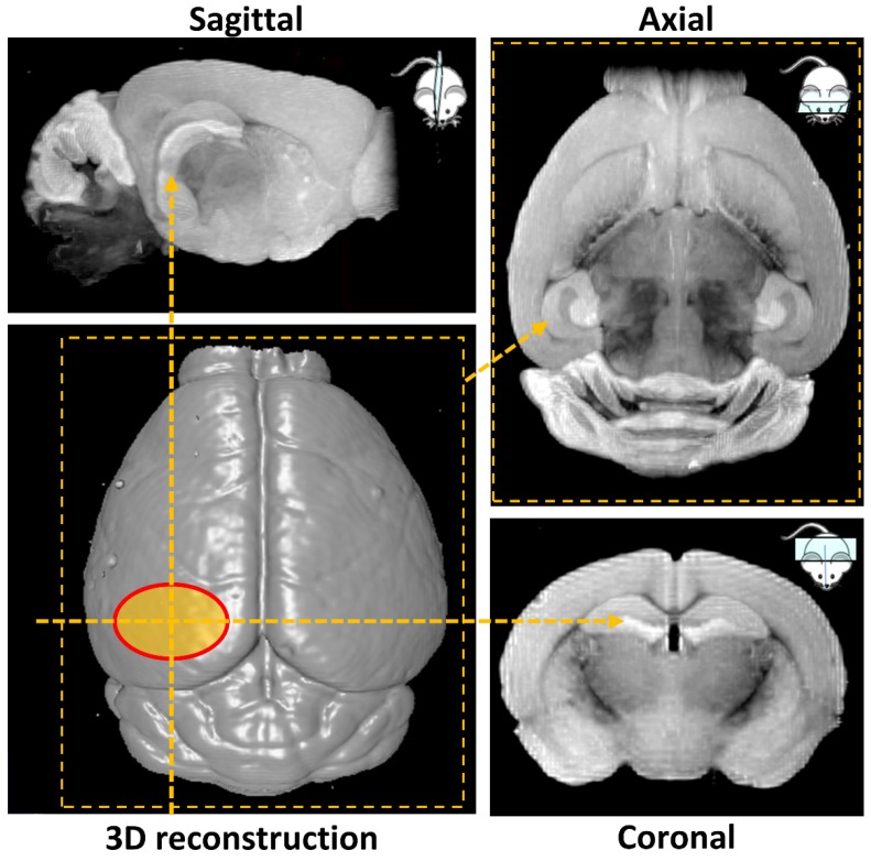
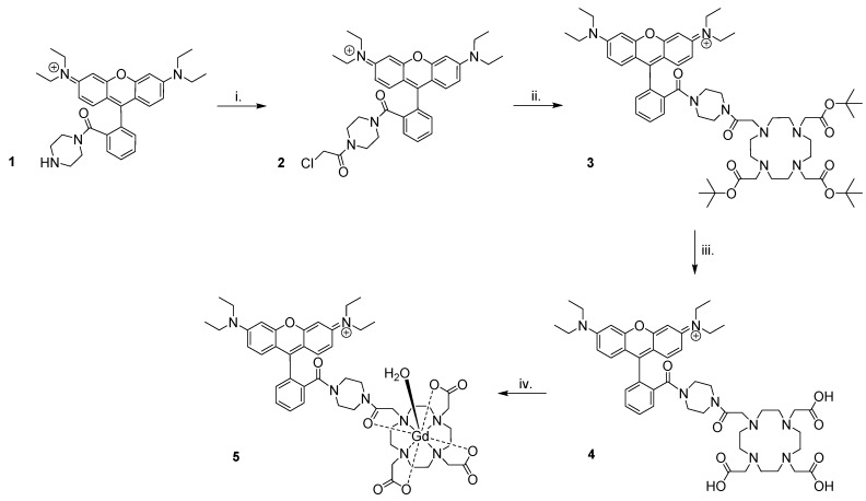
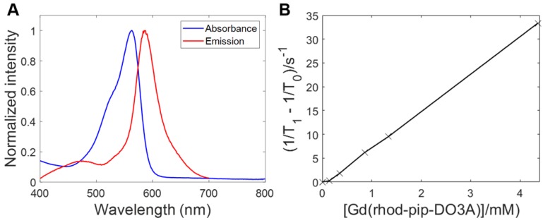
Gadolinium-based magnetic resonance imaging contrast agents can provide information regarding neuronal function, provided that these agents can cross the neuronal cell membrane. Such contrast agents are normally restricted to extracellular domains, however, by attaching cationic fluorescent dyes, they can be made cell-permeable and allow for both optical and magnetic resonance detection. To reach neurons, these agents also need to cross the blood-brain barrier. Focused ultrasound combined with microbubbles has been shown to enhance the permeability of this barrier, allowing molecules into the brain non-invasively, locally and transiently. The goal of this study was to investigate whether combining fluorescent rhodamine with a gadolinium complex would form a dual-modal contrast agent that could label neurons when delivered to the mouse brain with focused ultrasound and microbubbles. : Gadolinium complexes were combined with a fluorescent, cationic rhodamine unit to form probes with fluorescence and relaxivity properties suitable for applications. The left hemisphere of female C57bl/6 mice (8-10 weeks old; 19.07 ± 1.56 g; n = 16) was treated with ultrasound (centre frequency: 1 MHz, peak-negative pressure: 0.35 MPa, pulse length: 10 ms, repetition frequency: 0.5 Hz) while intravenously injecting SonoVue microbubbles and either the 1 kDa Gd(rhodamine-pip-DO3A) complex or a conventionally-used lysine-fixable Texas Red® 3 kDa dextran. The opposite right hemisphere was used as a non-treated control region. Brains were then extracted and either sectioned and imaged via fluorescence or confocal microscopy or imaged using a 9.4 T magnetic resonance imaging scanner. Brain slices were stained for neurons (NeuN), microglia (Iba1) and astrocytes (GFAP) to investigate the cellular localization of the probes. : Rhodamine fluorescence was detected in the left hemisphere of all ultrasound treated mice, while none was detected in the right control hemisphere. Cellular uptake of Gd(rhodamine-pip-DO3A) was observed in all the treated regions with a uniform distribution (coefficient of variation = 0.4 ± 0.05). Uptake was confirmed within neurons, whereas the probe did not co-localize with microglia and astrocytes. Compared to the dextran molecule, Gd(rhodamine-pip-DO3A) distributed more homogeneously and was less concentrated around blood vessels. Furthermore, the dextran molecule was found to accumulate unselectively in microglia as well as neurons, whereas our probe was only taken up by neurons. Gd(rhodamine-pip-DO3A) was detected via magnetic resonance imaging in similar regions to where fluorescence was detected. : We have introduced a method to image neurons with a dual-modal imaging agent delivered non-invasively and locally to the brain using focused ultrasound and microbubbles. When delivered to the mouse brain, the agent distributed homogeneously and was only uptaken by neurons; in contrast, conventionally used dextran distributed heterogeneously and was uptaken by microglia as well as neurons. This result indicates that our probe labels neurons without microglial involvement and in addition the probe was found to be detectable via both MRI and fluorescence. Labeling neurons with such dual-modal agents could facilitate the study of neuronal morphology and physiology using the advantages of both imaging modalities.
基于钆的磁共振成像造影剂能够提供有关神经元功能的信息,前提是这些造影剂能够穿过神经元细胞膜。然而,这类造影剂通常局限于细胞外区域,通过连接阳离子荧光染料,可使其具有细胞通透性,从而实现光学和磁共振检测。为了到达神经元,这些造影剂还需要穿过血脑屏障。聚焦超声联合微泡已被证明可增强该屏障的通透性,使分子能够以非侵入性、局部且短暂的方式进入大脑。本研究的目的是探究将荧光罗丹明与钆配合物相结合,是否能形成一种双模态造影剂,在通过聚焦超声和微泡递送至小鼠大脑时能够标记神经元。:将钆配合物与荧光阳离子罗丹明单元相结合,形成具有适合应用的荧光和弛豫特性的探针。对雌性C57bl/6小鼠(8 - 10周龄;19.07±1.56克;n = 16)的左半球进行超声处理(中心频率:1兆赫,负峰值压力:0.35兆帕,脉冲长度:10毫秒,重复频率:0.5赫兹),同时静脉注射声诺维微泡以及1千道尔顿的钆(罗丹明 - 哌啶 - DO3A)配合物或传统使用的可赖氨酸固定的德克萨斯红® 3千道尔顿葡聚糖。相对的右半球用作未处理的对照区域。然后取出大脑,要么通过荧光或共聚焦显微镜进行切片成像,要么使用9.4特斯拉磁共振成像扫描仪进行成像。对脑切片进行神经元(NeuN)、小胶质细胞(Iba1)和星形胶质细胞(GFAP)染色,以研究探针的细胞定位。:在所有接受超声处理的小鼠的左半球均检测到罗丹明荧光,而在右对照半球未检测到。在所有处理区域均观察到钆(罗丹明 - 哌啶 - DO3A)的细胞摄取,且分布均匀(变异系数 = 0.4±0.05)。在神经元内证实有摄取,而该探针未与小胶质细胞和星形胶质细胞共定位。与葡聚糖分子相比,钆(罗丹明 - 哌啶 - DO3A)分布更均匀,且在血管周围的聚集较少。此外,发现葡聚糖分子在小胶质细胞以及神经元中无选择性地积累,而我们的探针仅被神经元摄取。通过磁共振成像在与检测到荧光的类似区域检测到了钆(罗丹明 - 哌啶 - DO3A)。:我们引入了一种方法,使用聚焦超声和微泡将双模态成像剂以非侵入性和局部方式递送至大脑来对神经元进行成像。当递送至小鼠大脑时,该造影剂分布均匀且仅被神经元摄取;相比之下,传统使用的葡聚糖分布不均匀且被小胶质细胞以及神经元摄取。这一结果表明我们的探针在无小胶质细胞参与的情况下标记神经元,此外还发现该探针可通过磁共振成像和荧光检测到。使用此类双模态试剂标记神经元可利用两种成像方式的优势促进对神经元形态和生理学的研究。