The Third Hospital of Shanxi Medical University, Shanxi Bethune Hospital, Shanxi Academy of Medical Sciences, Taiyuan, 030032, People's Republic of China.
Int J Nanomedicine. 2022 Jun 30;17:2883-2890. doi: 10.2147/IJN.S367721. eCollection 2022.
Tumor derived cellular microvesicles (TDMVs), as excellent drug delivery vehicles in vivo, play an important role in the treatment of cancers. However, it is difficult to obtain intuitional biodistribution behavior and internalization mechanisms of TDMVs in vivo. Thus, it is very urgent and important to establish a stable and reliable visualization technology to track the biological behavior and function of TDMVs. As an endogenous biopolymer, melanin possesses natural biocompatibility and biodegradability, and various biological imaging could be realized by modifying it. Therefore, melanin-based nanoparticles are excellent candidates for in vivo visualization of TDMVs.
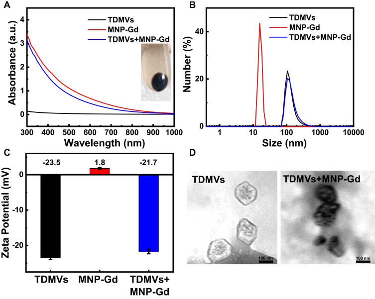
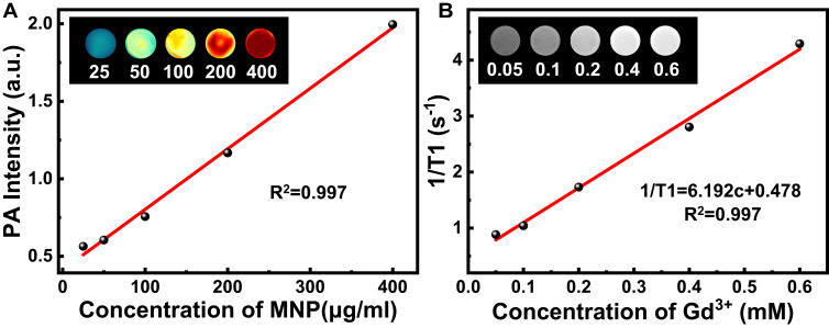
In this work, the biodistribution and metabolic behavior of TDMVs were visualized by dual-modality imaging with PAI and MRI after incubation with gadolinium ion-chelated melanin nanoparticles.
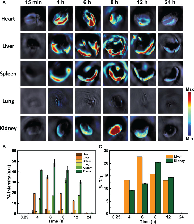
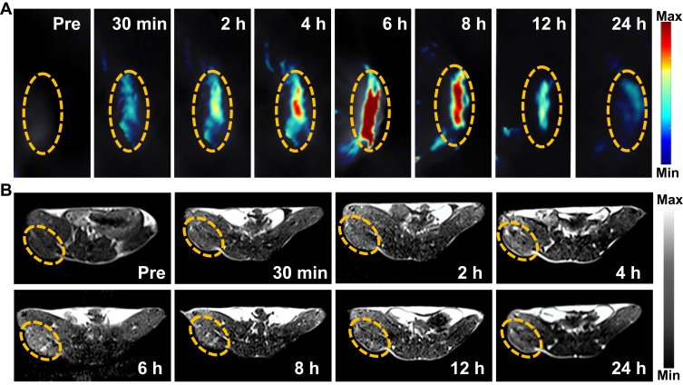
In this study, MRI and PAI dual-modality imaging of the in vivo distribution behavior of TDMVs was achieved with the help of MNP-Gd. The good targeting ability of TDMVs at the homologous tumor site was observed, and their distribution and metabolism behavior in the whole body were studied at the meantime. The results indicated that TDMVs preferentially accumulated in syngeneic tumor sites and liver regions after intravenous injection and were eventually metabolized by the kidneys over time.
This work proposed a novel dual-modal imaging strategy for the visualization of TDMVs, which is of great significance for further understanding the biological mechanisms of extracellular vesicles.
肿瘤衍生的细胞外囊泡(TDMVs)作为体内优秀的药物递送载体,在癌症治疗中发挥着重要作用。然而,在体内获得 TDMVs 的直观分布行为和内化机制是困难的。因此,建立一种稳定可靠的可视化技术来跟踪 TDMVs 的生物学行为和功能非常紧迫和重要。黑色素作为一种内源性生物聚合物,具有天然的生物相容性和生物降解性,通过修饰它可以实现各种生物成像。因此,基于黑色素的纳米颗粒是体内可视化 TDMVs 的优秀候选物。
在这项工作中,通过用镧系元素离子螯合黑色素纳米颗粒孵育后,进行 PAI 和 MRI 的双模式成像,可视化 TDMVs 的分布和代谢行为。
在这项研究中,借助 MNP-Gd 实现了 TDMVs 在体内分布行为的 MRI 和 PAI 双模式成像。观察到 TDMVs 在同源肿瘤部位具有良好的靶向能力,并同时研究了它们在全身的分布和代谢行为。结果表明,TDMVs 静脉注射后优先聚集在同基因肿瘤部位和肝脏区域,并随着时间的推移最终被肾脏代谢。
本工作提出了一种用于可视化 TDMVs 的新型双模式成像策略,对进一步了解细胞外囊泡的生物学机制具有重要意义。