Molecular Pathology of Cancer Group, Navarrabiomed, Complejo Hospitalario de Navarra (CHN), Universidad Pública de Navarra (UPNA), Instituto de Investigación Sanitaria de Navarra (IdiSNA), Irunlarrea 3, 31008 Pamplona, Spain.
Proteored-ISCIII, Proteomics Unit, Navarrabiomed, Complejo Hospitalario de Navarra (CHN), Universidad Pública de Navarra (UPNA), Instituto de Investigación Sanitaria de Navarra (IdiSNA), Irunlarrea 3, 31008 Pamplona, Spain.
Int J Mol Sci. 2020 Mar 19;21(6):2125. doi: 10.3390/ijms21062125.
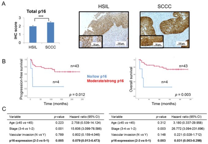
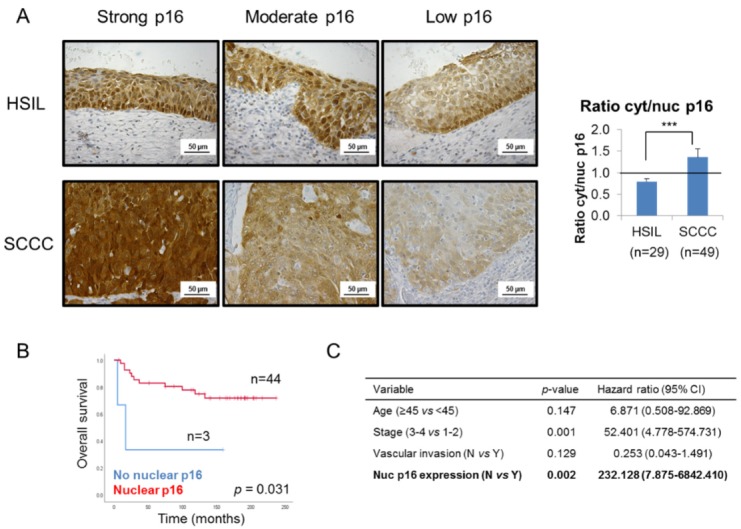
The tumor-suppressor protein p16 is paradoxically overexpressed in cervical cancer (CC). Despite its potential as a biomarker, its clinical value and the reasons for its failure in tumor suppression remain unclear. Our purpose was to determine p16 clinical and biological significance in CC. p16 expression pattern was examined by immunohistochemistry in 78 CC cases (high-grade squamous intraepithelial lesions (HSILs) and squamous cell carcinomas of the cervix -SCCCs). CC cell proliferation and invasion were monitored by real-time cell analysis and Transwell invasion assay, respectively. Cytoplasmic p16 interactors were identified from immunoprecipitated extracts by liquid chromatography-tandem mass spectrometry, and colocalization was confirmed by double-immunofluorescence. We observed that SCCCs showed significantly more cytoplasmic than nuclear p16 expression than HSILs. Importantly, nuclear p16 absence significantly predicted poor outcome in SCCC patients irrespective of other clinical parameters. Moreover, we demonstrated that cytoplasmic p16 interacted with CDK4 and other unreported proteins, such as BANF1, AKAP8 and AGTRAP, which could sequester p16 to avoid nuclear translocation, and then, impair its anti-tumor function. Our results suggest that the absence of nuclear p16 could be a diagnostic biomarker between HSIL and SCCC, and an independent prognostic biomarker in SCCC; and explain why p16 overexpression fails to stop CC growth.
抑癌蛋白 p16 在宫颈癌(CC)中表现出反常的过表达。尽管它具有作为生物标志物的潜力,但它在肿瘤抑制中的临床价值和失败的原因仍不清楚。我们的目的是确定 p16 在 CC 中的临床和生物学意义。通过免疫组织化学检测 78 例 CC 病例(高级别鳞状上皮内病变(HSIL)和宫颈鳞状细胞癌-SCCCs)中的 p16 表达模式。通过实时细胞分析和 Transwell 侵袭试验分别监测 CC 细胞的增殖和侵袭。通过液相色谱-串联质谱法从免疫沉淀提取物中鉴定细胞质 p16 相互作用蛋白,并通过双免疫荧光进行共定位验证。我们观察到 SCCCs 的细胞质 p16 表达明显多于 HSILs 的核表达。重要的是,无论其他临床参数如何,核 p16 缺失均显著预示 SCC 患者预后不良。此外,我们证明细胞质 p16 与 CDK4 和其他未报道的蛋白质相互作用,如 BANF1、AKAP8 和 AGTRAP,这些蛋白质可以将 p16 隔离以避免核易位,从而损害其抗肿瘤功能。我们的研究结果表明,核 p16 的缺失可能是 HSIL 和 SCC 之间的诊断生物标志物,也是 SCC 中的独立预后生物标志物;并解释了为什么 p16 过表达不能阻止 CC 生长。