Department of Radiology, Central Hospital of Wuhan, Tongji Medical College, Huazhong University of Science and Technology, Wuhan, China.
Cancer Center, Union Hospital, Tongji Medical College, Huazhong University of Science and Technology, Wuhan, China.
Korean J Radiol. 2020 May;21(5):541-544. doi: 10.3348/kjr.2020.0180. Epub 2020 Mar 20.
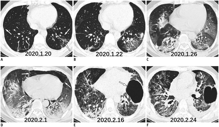
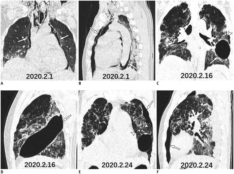
The coronavirus disease 2019 (COVID-19) pneumonia is a recent outbreak in mainland China and has rapidly spread to multiple countries worldwide. Pulmonary parenchymal opacities are often observed during chest radiography. Currently, few cases have reported the complications of severe COVID-19 pneumonia. We report a case where serial follow-up chest computed tomography revealed progression of pulmonary lesions into confluent bilateral consolidation with lower lung predominance, thereby confirming COVID-19 pneumonia. Furthermore, complications such as mediastinal emphysema, giant bulla, and pneumothorax were also observed during the course of the disease.
新型冠状病毒肺炎(COVID-19)是中国内地近期暴发的一种疾病,已迅速蔓延至全球多个国家。胸部 X 线摄影常可见肺部实质混浊。目前,鲜有报道严重 COVID-19 肺炎的并发症。我们报告了一例病例,连续随访的胸部 CT 显示肺部病变进展为双侧融合性实变,以下肺为主,从而确诊为 COVID-19 肺炎。此外,在疾病过程中还观察到了纵隔气肿、巨大肺大疱和气胸等并发症。