Department of Psychiatry and Psychotherapy, University of Regensburg, Regensburg, Germany.
Faculty of Psychology and Education Sciences, University of Coimbra, Coimbra, Portugal.
Sci Rep. 2020 Mar 26;10(1):5536. doi: 10.1038/s41598-020-62590-y.
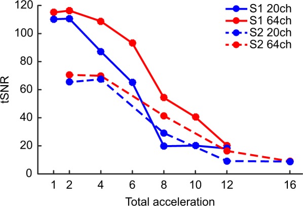
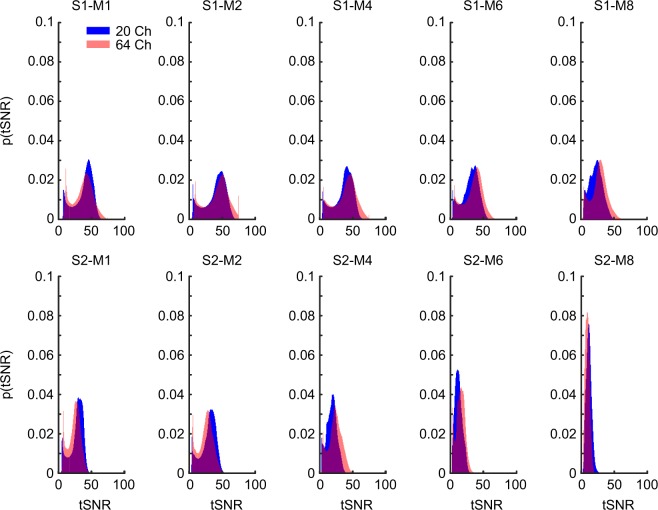
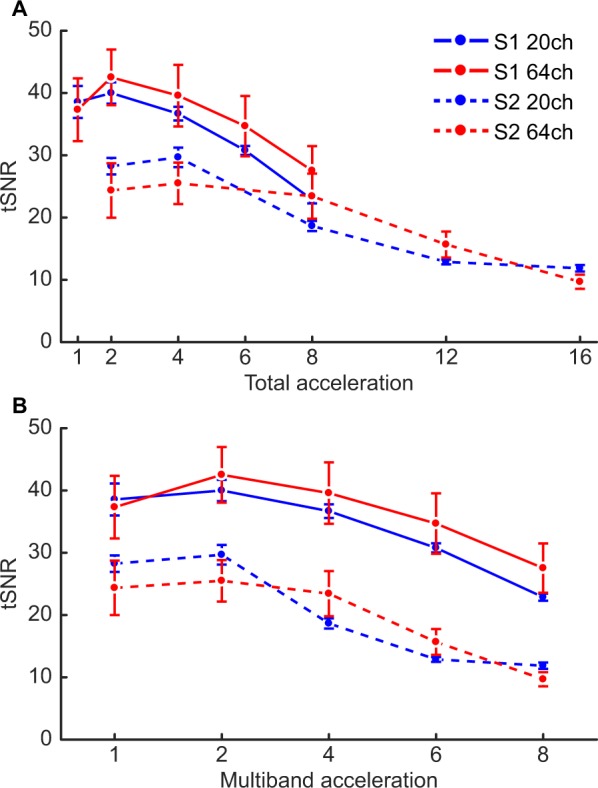
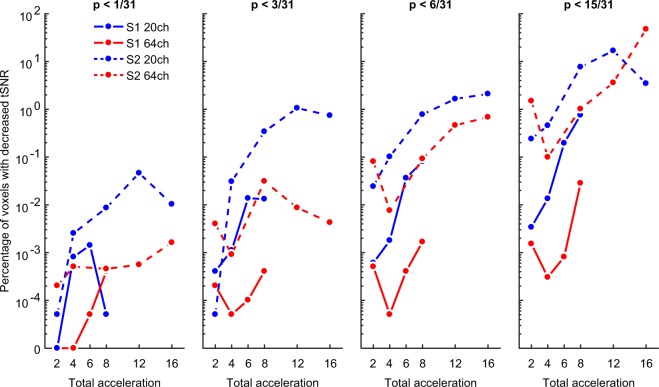
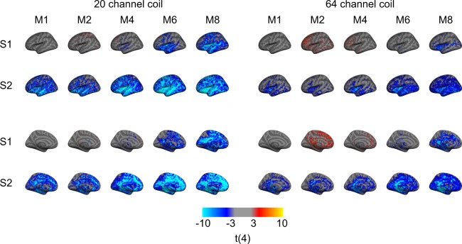
Echo-planar imaging (EPI) is the most common method of functional MRI for acquiring the blood oxygenation level-dependent (BOLD) contrast, allowing the acquisition of an entire brain volume within seconds. However, because imaging protocols are limited by hardware (e.g., fast gradient switching), researchers must compromise between spatial resolution, temporal resolution, or whole-brain coverage. Earlier attempts to circumvent this problem included developing protocols in which slices of a volume were acquired faster (i.e., in-plane acceleration (S)) or simultaneously (i.e., multislice acceleration (M)). However, applying acceleration methods can lead to a reduction in the temporal signal-to-noise ratio (tSNR): a critical measure of signal stability over time. Using a 20- and 64-channel receiver coil, we show that enabling S-acceleration consistently yielded a substantial decrease in tSNR, regardless of the receiver coil, whereas M-acceleration yielded less pronounced tSNR decrease. Moreover, tSNR losses tended to occur in temporal, insular, and medial brain regions and were more noticeable with the 20-channel coil, while with the 64-channel coil, the tSNR in lateral frontoparietal regions remained relatively stable up to six-fold M-acceleration producing comparable tSNR to that of no acceleration. Such methodological explorations can guide researchers and clinicians in optimizing imaging protocols depending on the brain regions under investigation.
平面回波成像(EPI)是采集血氧水平依赖(BOLD)对比功能磁共振成像最常用的方法,允许在几秒钟内采集整个大脑体积。然而,由于成像协议受到硬件(例如快速梯度切换)的限制,研究人员必须在空间分辨率、时间分辨率或全脑覆盖之间进行权衡。早期为解决此问题的尝试包括开发更快采集体积切片的协议(即,平面内加速(S))或同时采集(即,多切片加速(M))。然而,应用加速方法可能会导致时间信号噪声比(tSNR)降低:这是衡量信号随时间稳定性的关键指标。我们使用 20 通道和 64 通道接收线圈显示,无论接收线圈如何,S 加速始终会导致 tSNR 大幅降低,而 M 加速则导致 tSNR 降低不太明显。此外,tSNR 损失往往发生在时间、岛叶和内侧脑区,并且在 20 通道线圈中更为明显,而在 64 通道线圈中,直到六倍的 M 加速产生与无加速相当的 tSNR 之前,侧额顶叶区域的 tSNR 仍然相对稳定。这种方法学探索可以指导研究人员和临床医生根据研究的脑区优化成像协议。