Schmitt Tina, Rieger Jochem W
Neuroimaging Unit, School of Medicine and Health Sciences, Carl von Ossietzky Universität Oldenburg, Oldenburg, Germany.
Department of Psychology, School of Medicine and Health Sciences, Carl von Ossietzky Universität Oldenburg, Oldenburg, Germany.
Front Neurosci. 2021 Oct 29;15:735290. doi: 10.3389/fnins.2021.735290. eCollection 2021.
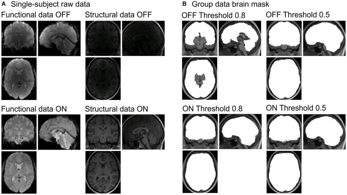
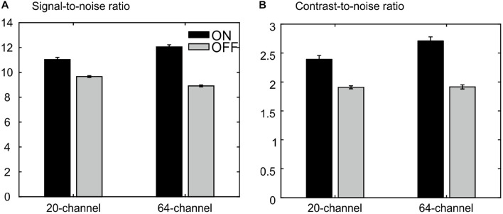
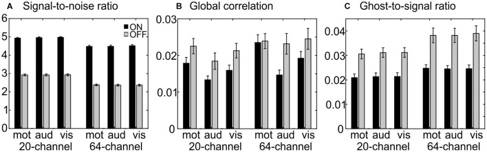
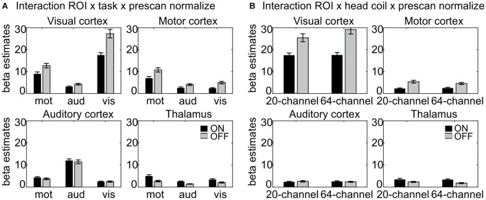
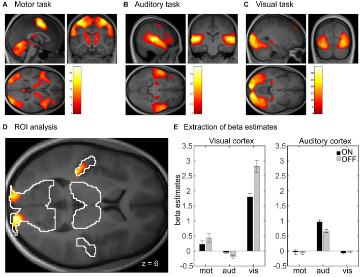
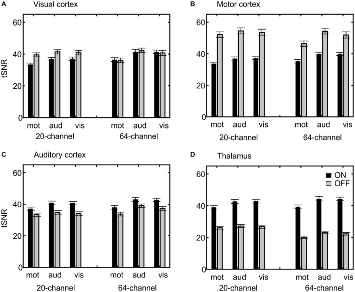
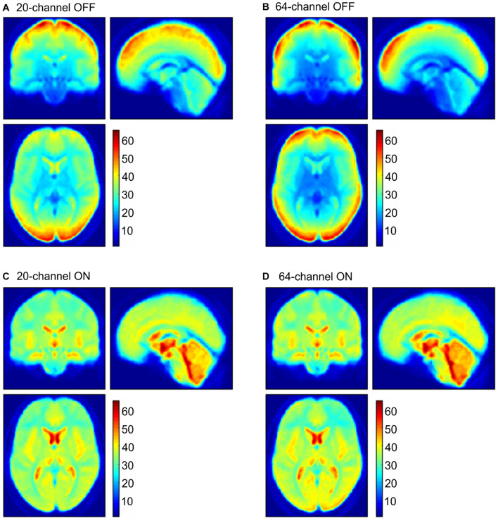
The performance of MRI head coils together with the influence of the prescan normalize filter in different brain regions was evaluated. Functional and structural data were recorded from 26 participants performing motor, auditory, and visual tasks in different conditions: with the 20- and 64-channel Siemens head/neck coil and the prescan normalize filter turned ON or OFF. Data were analyzed with the MRIQC tool to evaluate data quality differences. The functional data were statistically evaluated by comparison of the β estimates and the time-course signal-to-noise ratio (tSNR) in four regions of interest, i.e., the auditory, visual, and motor cortices and the thalamus. The MRIQC tool indicated a better data quality for both functional and structural data with the prescan normalize filter, with an advantage for the 20-channel head coil in functional data and an advantage for the 64-channel head coil in structural measurements. Nevertheless, recommendations for the functional data regarding choice of head coils and prescan normalize filter depend on the brain regions of interest. Higher β estimates and tSNR values occurred in the auditory cortex and thalamus with the prescan normalize filter, whereas the contrary was true for the visual and motor cortices. Due to higher β estimates in the visual cortex in the 64-channel head coil, this head coil is recommended for studies investigating the visual cortex. For most of the research questions, the 20-channel head coil is better suited for functional experiments, with the prescan normalize filter, especially when investigating deep brain areas. For anatomical studies, the 64-channel head coil seemed to be the better choice.
评估了MRI头部线圈的性能以及预扫描归一化滤波器在不同脑区的影响。记录了26名参与者在不同条件下执行运动、听觉和视觉任务时的功能和结构数据:使用20通道和64通道西门子头/颈线圈,且预扫描归一化滤波器开启或关闭。使用MRIQC工具分析数据以评估数据质量差异。通过比较四个感兴趣区域(即听觉、视觉和运动皮层以及丘脑)的β估计值和时间进程信噪比(tSNR)对功能数据进行统计学评估。MRIQC工具表明,使用预扫描归一化滤波器时,功能和结构数据的质量均更好,在功能数据方面20通道头部线圈具有优势,在结构测量方面64通道头部线圈具有优势。然而,关于头部线圈和预扫描归一化滤波器选择的功能数据建议取决于感兴趣的脑区。使用预扫描归一化滤波器时,听觉皮层和丘脑中出现了更高的β估计值和tSNR值,而视觉和运动皮层则相反。由于64通道头部线圈在视觉皮层中的β估计值更高,因此推荐该头部线圈用于研究视觉皮层。对于大多数研究问题,20通道头部线圈更适合进行功能实验,使用预扫描归一化滤波器,尤其是在研究深部脑区时。对于解剖学研究,64通道头部线圈似乎是更好的选择。