Franco-Barraza Janusz, Raghavan Kristopher S, Luong Tiffany, Cukierman Edna
Cancer Biology, The Martin and Concetta Greenberg Pancreatic Cancer Institute, Fox Chase Cancer Center, Philadelphia, PA, United States.
Cancer Biology, The Martin and Concetta Greenberg Pancreatic Cancer Institute, Fox Chase Cancer Center, Philadelphia, PA, United States; College of Medicine, Drexel University, Philadelphia, PA, United States.
Methods Cell Biol. 2020;156:109-160. doi: 10.1016/bs.mcb.2019.11.014. Epub 2020 Jan 21.
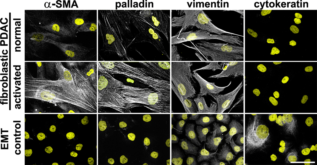
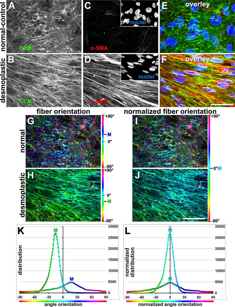
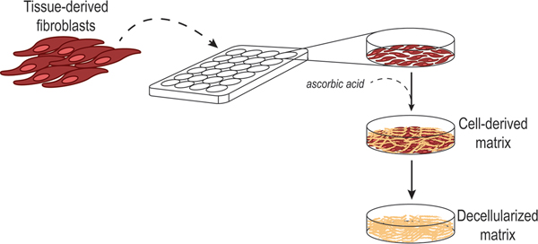
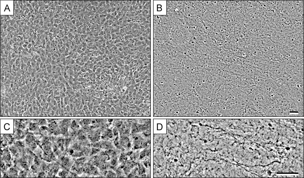
Three-dimensional (3D) culturing models, replicating in vivo tissue microenvironments that incorporate native extracellular matrix (ECM), have revolutionized the cell biology field. Fibroblastic cells generate lattices of interstitial ECM proteins. Cell interactions with ECMs and with molecules sequestered/stored within these are crucial for tissue development and homeostasis maintenance. Hence, ECMs provide cells with biochemical and biomechanical cues to support and locally control cell function. Further, dynamic changes in ECMs, and in cell-ECM interactions, partake in growth, development, and temporary occurrences such as acute wound healing. Notably, dysregulation in ECMs and fibroblasts could be important triggers and modulators of pathological events such as developmental defects, and diseases associated with fibrosis and chronic inflammation such as cancer. Studying the type of fibroblastic cells producing these matrices and how alterations to these cells enable changes in ECMs are of paramount importance. This chapter provides a step-by-step method for producing multilayered (e.g., 3D) fibroblastic cell-derived matrices (fCDM). Methods also include means to assess ECM topography and other cellular traits, indicative of fibroblastic functional statuses, like naïve/normal vs. inflammatory and/or myofibroblastic. For these, protocols include indications for isolating normal and diseased fibroblasts (i.e., cancer-associated fibroblasts known as CAFs). Protocols also include means for conducting microscopy assessments, querying whether fibroblasts present with fCDM-dependent normal or CAF phenotypes. These are supported by discrete semi-quantitative digital imaging analyses, providing some imaging processing advice. Additionally, protocols include descriptions for effective fCDM decellularization, which renders cellular debris-free patho/physiological in vivo-like scaffolds, suitable as 3D substrates for subsequent cell culturing.
三维(3D)培养模型能够复制包含天然细胞外基质(ECM)的体内组织微环境,彻底改变了细胞生物学领域。成纤维细胞生成间质ECM蛋白的晶格。细胞与ECM以及与隔离/存储在其中的分子之间的相互作用对于组织发育和内环境稳态的维持至关重要。因此,ECM为细胞提供生化和生物力学线索,以支持和局部控制细胞功能。此外,ECM以及细胞与ECM相互作用的动态变化参与生长、发育以及诸如急性伤口愈合等临时事件。值得注意的是,ECM和成纤维细胞的失调可能是诸如发育缺陷以及与纤维化和慢性炎症相关的疾病(如癌症)等病理事件的重要触发因素和调节因素。研究产生这些基质的成纤维细胞类型以及这些细胞的改变如何导致ECM的变化至关重要。本章提供了一种逐步生成多层(例如3D)成纤维细胞衍生基质(fCDM)的方法。方法还包括评估ECM拓扑结构和其他细胞特征的手段,这些特征指示成纤维细胞的功能状态,如幼稚/正常与炎症和/或肌成纤维细胞状态。为此,方案包括分离正常和患病成纤维细胞(即癌症相关成纤维细胞,称为CAF)的指示。方案还包括进行显微镜评估的手段,以查询成纤维细胞是否呈现依赖fCDM的正常或CAF表型。这些由离散的半定量数字成像分析支持,并提供了一些成像处理建议。此外,方案还包括有效fCDM去细胞化的描述,这可产生无细胞碎片的病理/生理体内样支架,适合作为后续细胞培养的3D底物。