U-Vet Animal Hospital, Faculty of Veterinary and Agricultural Sciences, University of Melbourne, 250 Princes Highway, Werribee, Victoria, 3030, Australia.
Statistical Consulting Centre, University of Melbourne, 139 Barry Street, Carlton, Melbourne, Victoria, 3053, Australia.
BMC Vet Res. 2020 Mar 30;16(1):104. doi: 10.1186/s12917-020-02327-1.
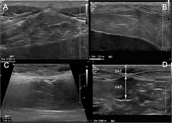
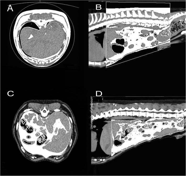
Adipose tissue may have different metabolic and endocrine functions depending on the region of the body in which it is located. While visceral or intra-abdominal fat has been found to contribute to leptin concentrations, insulin resistance and obesity-related diseases, there are only a few imaging studies documenting the preferential distribution of body fat to either the intra-abdominal or subcutaneous compartments in dogs. This study aimed to determine if CT-measured abdominal fat distributed preferentially to the visceral space (V) relative to the subcutaneous space (SQ), with increasing DXA-determined total body fat percentage; and if ultrasound measurements of the ventral midline subcutaneous (SAT) and visceral adipose thickness (VAT) can be used to estimate the distribution of fat to the subcutaneous and visceral abdominal spaces, in a sample of 22 dogs with variable body condition.
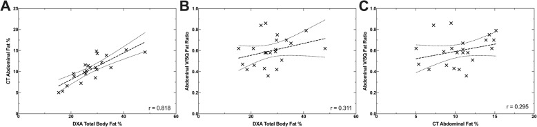
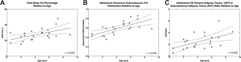
Multivariate analysis showed no statistically significant correlation between visceral to subcutaneous fat ratio (V/SQ) and increasing total body fat percentage (β = - 0.07, p = 0.733), but strong correlation with age (β = 0.71 p = 0.002). A substantial amount of variation for the ultrasound visceral adipose thickness to subcutaneous fat thickness (VAT/SAT) could be explained by both CT V/SQ and sex (R = 0.477, p = 0.001), with female dogs having significant lower VAT/SAT ratios compared to the male dogs (p = 0.047). The ultrasound fat measurements appeared moderately reliable, but a larger sample number is required to confirm this.
The findings suggest that dogs with a relatively healthy to slightly overweight body condition score, distribute fat relatively similarly between their peritoneal (visceral) and subcutaneous abdominal compartments with increasing total body fat percentage. However, there was increased fat distribution to the peritoneal space relative to the subcutaneous space with increasing age. Further, abdominal ultrasound may be useful in estimating the ratio of fat distribution to both the abdominal visceral and subcutaneous spaces.
脂肪组织可能具有不同的代谢和内分泌功能,具体取决于其所在的身体部位。虽然内脏或腹腔内脂肪已被发现会导致瘦素浓度、胰岛素抵抗和与肥胖相关的疾病增加,但只有少数影像学研究记录了狗体内脂肪优先分布于腹腔内或皮下隔室。本研究旨在确定 CT 测量的腹部脂肪相对于皮下隔室(SQ)是否优先分布于内脏空间(V),随着 DXA 确定的总身体脂肪百分比的增加;以及在一个 22 只身体状况不同的狗的样本中,是否可以使用超声测量腹中线皮下(SAT)和内脏脂肪厚度(VAT)来估计脂肪分布到皮下和内脏腹部空间。
多变量分析显示,内脏与皮下脂肪比率(V/SQ)与总身体脂肪百分比的增加之间没有统计学上的显著相关性(β=−0.07,p=0.733),但与年龄有很强的相关性(β=0.71,p=0.002)。超声内脏脂肪厚度与皮下脂肪厚度(VAT/SAT)的大量变异可以通过 CT V/SQ 和性别来解释(R=0.477,p=0.001),与雄性狗相比,雌性狗的 VAT/SAT 比值显著较低(p=0.047)。超声脂肪测量结果显示出中度可靠性,但需要更大的样本量来证实这一点。
研究结果表明,在身体状况相对健康到略超重的范围内,随着总身体脂肪百分比的增加,狗的腹膜(内脏)和皮下腹部隔室之间的脂肪分布相对相似。然而,随着年龄的增长,脂肪在腹膜空间的分布相对于皮下空间增加。此外,腹部超声可能有助于估计腹部内脏和皮下脂肪分布的比例。