Department of Orthopaedic Surgery, Regensburg University Medical Centre, Asklepios Klinikum Bad Abbach, Kaiser-Karl-V-Allee 3, 93077, Bad Abbach, Germany.
Arch Orthop Trauma Surg. 2020 Jul;140(7):933-940. doi: 10.1007/s00402-020-03422-6. Epub 2020 Mar 30.
Valgus deformity presents a particular challenge in total knee arthroplasty. This condition regularly leads to contractures of the lateral capsular ligament complex and to overstretching of the medial ligamentous complex. Reconstruction of the knee joint kinematics and anatomy often requires lateral release. However, data on how such release weakens the stability of the knee are missing in the literature. This study investigated the effects of sequential lateral release on the collateral stability of the ligament complex of the knee in vitro.
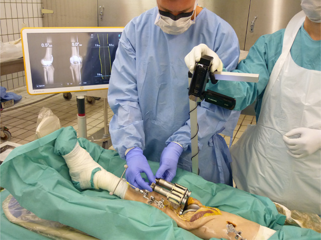
Ten knee prostheses were implanted in 10 healthy cadaveric knee joints using a navigation device. Soft tissue lateral release consisted of five release steps, and stiffness and stability were determined at 0, 30, 60 and 90° flexion after each step.
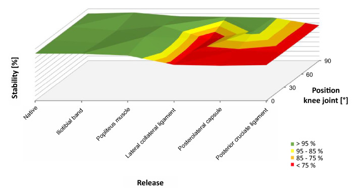
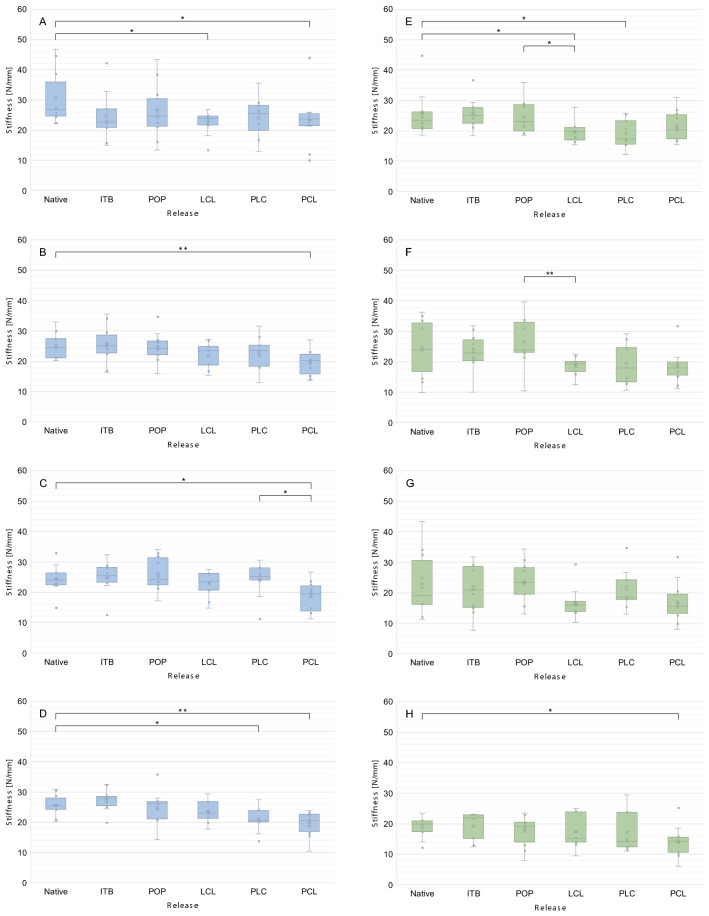
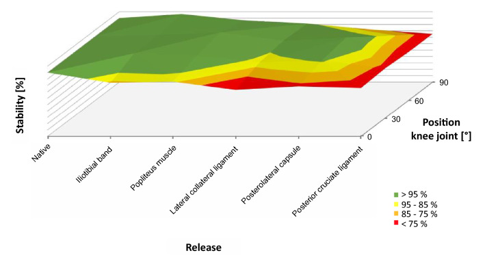
Soft tissue lateral release increasingly weakened the ligament complex of the lateral compartment. Because of the large muscular parts, the release of the iliotibial band and the M. popliteus had little effect on the stability of the lateral and medial compartment, but release of the lateral ligament significantly decreased the stability in the lateral compartment over the entire range of motion. Stability in the medial compartment was hardly affected. Conversely, further release of the posterolateral capsule and the posterior cruciate ligament led to the loss of stability in the lateral compartment only in deep flexion, whereas stability decreased significantly in the medial compartment.
Our study shows for the first time the association between sequential lateral release and stability of the ligamentous complex of the knee. To maintain the stability, knee surgeons should avoid releasing the entire lateral collateral ligament, which would significantly decrease stability in the lateral compartment.
在全膝关节置换术中,外翻畸形是一个特别具有挑战性的问题。这种情况通常会导致外侧囊韧带复合体挛缩和内侧韧带复合体过度拉伸。为了重建膝关节运动学和解剖结构,通常需要进行外侧松解。然而,文献中缺乏关于这种松解如何削弱膝关节稳定性的数据。本研究在体外研究了连续外侧松解对膝关节韧带复合体外侧稳定的影响。
使用导航设备将 10 个膝关节假体植入 10 个健康的尸体膝关节中。软组织外侧松解包括五个松解步骤,在每个步骤后,在 0、30、60 和 90°屈曲时测量韧带复合体的刚度和稳定性。
软组织外侧松解逐渐削弱了外侧间室的韧带复合体。由于肌肉部分较大,髂胫束和腓肠肌的释放对外侧和内侧间室的稳定性几乎没有影响,但外侧韧带的释放显著降低了整个运动范围内外侧间室的稳定性。内侧间室的稳定性几乎不受影响。相反,后外侧囊和后交叉韧带的进一步释放仅在深度屈曲时导致外侧间室的稳定性丧失,而内侧间室的稳定性则显著下降。
我们的研究首次显示了连续外侧松解与膝关节韧带复合体稳定性之间的关联。为了保持稳定性,膝关节外科医生应避免松解整个外侧副韧带,因为这会显著降低外侧间室的稳定性。