• 文献检索
  • 文档翻译
  • 深度研究
  • 学术资讯
  • Suppr Zotero 插件Zotero 插件
  • 邀请有礼
  • 套餐&价格
  • 历史记录
应用&插件
Suppr Zotero 插件Zotero 插件浏览器插件Mac 客户端Windows 客户端微信小程序
定价
高级版会员购买积分包购买API积分包
服务
文献检索文档翻译深度研究API 文档MCP 服务
关于我们
关于 Suppr公司介绍联系我们用户协议隐私条款
关注我们

Suppr 超能文献

核心技术专利:CN118964589B侵权必究
粤ICP备2023148730 号-1Suppr @ 2026

文献检索

告别复杂PubMed语法,用中文像聊天一样搜索,搜遍4000万医学文献。AI智能推荐,让科研检索更轻松。

立即免费搜索

文件翻译

保留排版,准确专业,支持PDF/Word/PPT等文件格式,支持 12+语言互译。

免费翻译文档

深度研究

AI帮你快速写综述,25分钟生成高质量综述,智能提取关键信息,辅助科研写作。

立即免费体验

膝关节后外侧角的关节镜解剖:解剖关系和关节镜入路。

Arthroscopic anatomy of the posterolateral corner of the knee: anatomic relations and arthroscopic approaches.

机构信息

Department of Trauma and Orthopaedic Surgery, University Medical Center Hamburg-Eppendorf, Martinistrasse 52, 20246, Hamburg, Germany.

Department of Orthopaedic and Trauma Surgery, University Medical Center Schleswig-Holstein, Kiel, Germany.

出版信息

Arch Orthop Trauma Surg. 2022 Mar;142(3):443-453. doi: 10.1007/s00402-021-03864-6. Epub 2021 Mar 22.

DOI:10.1007/s00402-021-03864-6
PMID:33751186
原文链接:https://pmc.ncbi.nlm.nih.gov/articles/PMC8843911/
Abstract

INTRODUCTION

Although open-surgical techniques for the reconstruction of the posterolateral corner (PLC) are well established, the use of arthroscopic procedures has recently increased. When compared with open surgical preparation, arthroscopic orientation in the PLC is challenging and anatomic relations may not be familiar. Nevertheless, a profound knowledge of anatomic key structures and possible structures at risk as well as technical variations of arthroscopic approaches are mandatory to allow a precise and safe surgical intervention.

MATERIALS AND METHODS

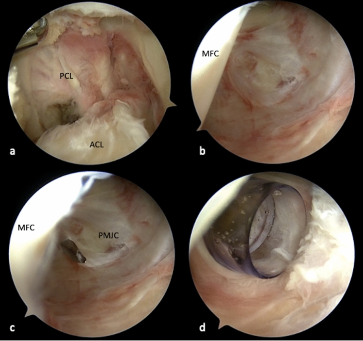
In a cadaveric video demonstration, an anterolateral (AL), anteromedial (AM), posteromedial (PM) and posterolateral (PL) portal, as well as a transseptal approach (TSA) were developed. Key structures of the PLC were defined and sequentially exposed during posterolateral arthroscopy. Finally, anatomic relations of all key structures were demonstrated.

RESULTS

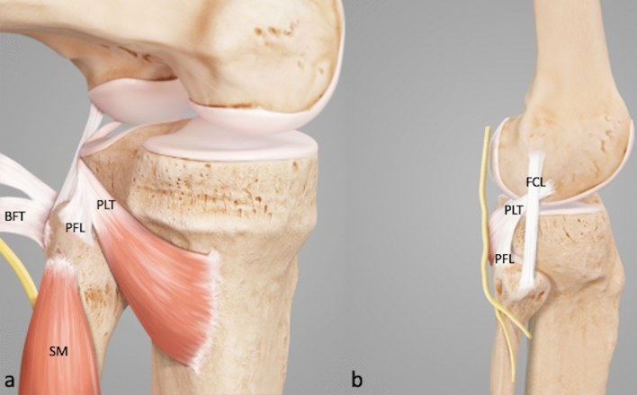
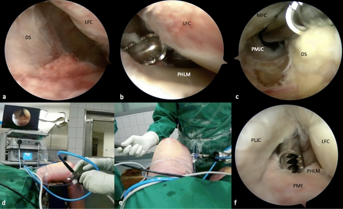
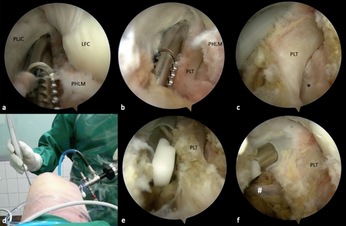
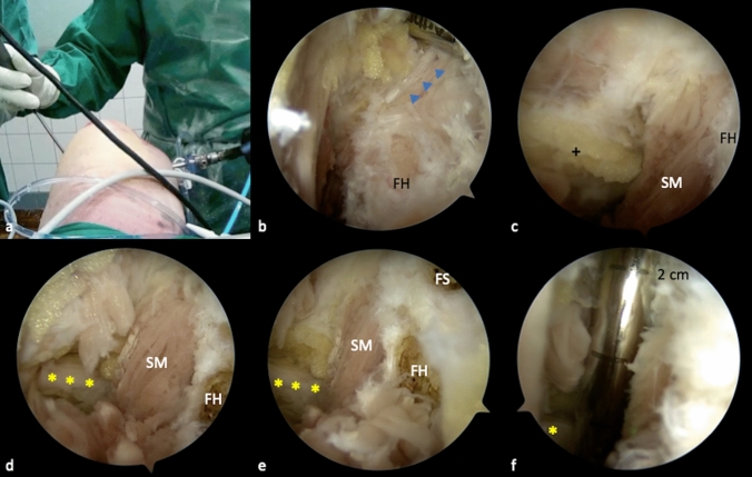
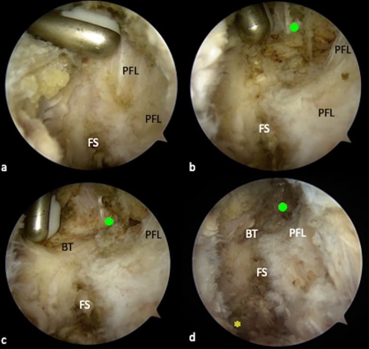
All key structures of the PLC can be visualized during arthroscopy. Thereby, careful portal placement is crucial in order to allow an effective exposure. Two alternatives of the TSA were described, depending on the region of interest. The peroneal nerve can be visualized dorsal to the biceps femoris tendon (BT), lateral to the soleus muscle (SM) and about 3 cm distal to the fibular styloid (FS). The distal attachment of the fibular collateral ligament (FCL) can be exposed on the lateral side of the fibular head (FH). The fibular attachment of the popliteofibular ligament (PFL) is exposed at the tip of the FS.

CONCLUSION

Arthroscopy of the posterolateral recessus allows full visualization of all key structures of the posterolateral corner, which provides the basis for anatomic and safe drill channel placement in PLC reconstruction. A sufficient exposure of relevant anatomic landmarks and precise portal preparation reduce the risk of iatrogenic vascular and peroneal nerve injury.

摘要

简介

尽管开放手术技术已广泛应用于后外侧角(PLC)的重建,但关节镜技术的应用近来有所增加。与开放手术准备相比,PLC 关节镜定位具有挑战性,解剖关系可能不熟悉。然而,为了实现精确和安全的手术干预,必须深入了解解剖关键结构和可能的风险结构,以及关节镜入路的技术变化。

材料与方法

在尸体视频演示中,开发了前外侧(AL)、前内侧(AM)、后内侧(PM)和后外侧(PL)入路,以及经隔入路(TSA)。定义了 PLC 的关键结构,并在进行后外侧关节镜检查时依次暴露这些结构。最后,演示了所有关键结构的解剖关系。

结果

所有 PLC 的关键结构均可在关节镜下可视化。因此,为了实现有效的暴露,仔细选择入路至关重要。根据感兴趣的区域,描述了 TSA 的两种替代方法。腓总神经可在比目鱼肌(SM)外侧、腓骨茎突(FS)远端 3 厘米处看到。外侧副韧带(LCL)的腓骨附着点可在腓骨头(FH)的外侧暴露。腘腓韧带(PFL)的腓骨附着点在 FS 的尖端暴露。

结论

后外侧隐窝关节镜检查可全面观察 PLC 的所有关键结构,为 PLC 重建中安全的钻孔通道定位提供解剖学基础。充分暴露相关解剖标志和精确的入路准备可降低医源性血管和腓总神经损伤的风险。

https://cdn.ncbi.nlm.nih.gov/pmc/blobs/5b6a/8843911/39e98df189dd/402_2021_3864_Fig6_HTML.jpg
https://cdn.ncbi.nlm.nih.gov/pmc/blobs/5b6a/8843911/ceeac6b81a8b/402_2021_3864_Fig1_HTML.jpg
https://cdn.ncbi.nlm.nih.gov/pmc/blobs/5b6a/8843911/890b1f1b72bd/402_2021_3864_Fig2_HTML.jpg
https://cdn.ncbi.nlm.nih.gov/pmc/blobs/5b6a/8843911/c87db92f40f5/402_2021_3864_Fig3_HTML.jpg
https://cdn.ncbi.nlm.nih.gov/pmc/blobs/5b6a/8843911/bcd352320b96/402_2021_3864_Fig4_HTML.jpg
https://cdn.ncbi.nlm.nih.gov/pmc/blobs/5b6a/8843911/96df6f1c172a/402_2021_3864_Fig5_HTML.jpg
https://cdn.ncbi.nlm.nih.gov/pmc/blobs/5b6a/8843911/39e98df189dd/402_2021_3864_Fig6_HTML.jpg

相似文献

1
Arthroscopic anatomy of the posterolateral corner of the knee: anatomic relations and arthroscopic approaches.膝关节后外侧角的关节镜解剖:解剖关系和关节镜入路。
Arch Orthop Trauma Surg. 2022 Mar;142(3):443-453. doi: 10.1007/s00402-021-03864-6. Epub 2021 Mar 22.
2
Morphology of the fibular insertion of the posterolateral corner and biceps femoris tendon.后外侧角和股二头肌肌腱腓骨附着处的形态学
Knee Surg Sports Traumatol Arthrosc. 2017 Jan;25(1):184-191. doi: 10.1007/s00167-016-4304-x. Epub 2016 Sep 12.
3
Posterolateral Rotatory Knee Instability-MRI Evaluation of Anatomic Landmarks for Tibial Drill Tunnel Placement in Open and Arthroscopic Popliteus Tendon Reconstruction.后外侧旋转性膝关节不稳——在开放性和关节镜下腘肌腱重建术中胫骨钻孔隧道置入解剖标志的MRI评估
J Knee Surg. 2019 Jul;32(7):667-672. doi: 10.1055/s-0038-1666832. Epub 2018 Jul 6.
4
Comparison of Arthroscopic versus Open Placement of the Fibular Tunnel in Posterolateral Corner Reconstruction.关节镜与开放腓骨隧道在后外侧角重建中的比较。
J Knee Surg. 2023 Jul;36(9):977-987. doi: 10.1055/s-0042-1748897. Epub 2022 Jul 7.
5
Posterolateral corner of the knee: a systematic literature review of current concepts of arthroscopic reconstruction.膝关节后外侧角:关节镜重建当前概念的系统文献回顾。
Arch Orthop Trauma Surg. 2020 Dec;140(12):2003-2012. doi: 10.1007/s00402-020-03607-z. Epub 2020 Sep 21.
6
Arcuate sign of posterolateral knee injuries: anatomic, radiographic, and MR imaging data related to patterns of injury.膝关节后外侧损伤的弓状征:与损伤模式相关的解剖学、放射学和磁共振成像数据
Skeletal Radiol. 2003 Nov;32(11):619-27. doi: 10.1007/s00256-003-0679-0. Epub 2003 Oct 1.
7
An All-Arthroscopic Technique for Complex Posterolateral Corner Reconstruction.一种用于复杂后外侧角重建的全关节镜技术。
Arthrosc Tech. 2019 Sep 11;8(9):e999-e1006. doi: 10.1016/j.eats.2019.05.010. eCollection 2019 Sep.
8
Can Anatomic Posterolateral Corner Reconstruction Using a Fibular Tunnel Restore Fibular Footprints of the Posterolateral Complex? A Cadaveric Study.采用腓骨隧道的解剖后外侧角重建能否恢复后外侧复合体的腓骨附着点?一项尸体研究。
Arthroscopy. 2020 May;36(5):1355-1362. doi: 10.1016/j.arthro.2019.11.099. Epub 2019 Dec 23.
9
Part I: An anatomic-based tunnel in the fibular head for posterolateral corner reconstruction using magnetic resonance imaging.第一部分:基于解剖结构的腓骨头隧道,用于使用磁共振成像进行后外侧角重建。
Knee Surg Sports Traumatol Arthrosc. 2015 Jul;23(7):1889-94. doi: 10.1007/s00167-014-3085-3. Epub 2014 Jun 7.
10
Lateral Knee Compartment Portals: A Cadaveric Study Defining a Posterolateral Viewing Safety Zone.外侧膝关节室入路:一项定义后外侧观察安全区的尸体研究。
Arthroscopy. 2018 Jul;34(7):2201-2206. doi: 10.1016/j.arthro.2018.02.006. Epub 2018 Apr 12.

引用本文的文献

1
Arthroscopic-Assisted Tibiofibular-Based Posterolateral Corner Knee Reconstruction.关节镜辅助下基于胫腓骨的膝关节后外侧角重建术
Arthrosc Tech. 2025 Jun 18;14(8):103698. doi: 10.1016/j.eats.2025.103698. eCollection 2025 Aug.
2
All-Arthroscopic Popliteus Tendon Reconstruction With Dual-Posterolateral Portal Technique.采用双后外侧入路技术的全关节镜下腘肌腱重建术。
Arthrosc Tech. 2025 Feb 13;14(6):103474. doi: 10.1016/j.eats.2025.103474. eCollection 2025 Jun.
3
Arthroscopic Arthrolysis, a Minimally Invasive Approach to Treat Arthrofibrosis of the Knee.

本文引用的文献

1
Anatomical evaluation of the femoral attachment of the posterior oblique ligament.后斜韧带股骨附着处的解剖评估。
Arch Orthop Trauma Surg. 2021 Jun;141(6):1035-1041. doi: 10.1007/s00402-020-03728-5. Epub 2021 Jan 8.
2
Posterolateral corner of the knee: a systematic literature review of current concepts of arthroscopic reconstruction.膝关节后外侧角:关节镜重建当前概念的系统文献回顾。
Arch Orthop Trauma Surg. 2020 Dec;140(12):2003-2012. doi: 10.1007/s00402-020-03607-z. Epub 2020 Sep 21.
3
The Popliteus Bypass provides superior biomechanical properties compared to the Larson technique in the reconstruction of combined posterolateral corner and posterior cruciate ligament injury.
关节镜下关节松解术,一种治疗膝关节纤维性关节僵直的微创方法。
Arthrosc Tech. 2025 Jan 31;14(5):103446. doi: 10.1016/j.eats.2025.103446. eCollection 2025 May.
4
Easy Arthroscopic Transseptal Approach of the Knee.膝关节简易关节镜经隔膜入路
Video J Sports Med. 2022 Jul 19;2(4):26350254221087290. doi: 10.1177/26350254221087290. eCollection 2022 Jul-Aug.
5
Exploring the Posterolateral Corner of the Knee Joint: A Detailed Review of Recent Literature.探索膝关节后外侧角:近期文献详细综述
J Clin Med. 2025 Feb 25;14(5):1549. doi: 10.3390/jcm14051549.
6
Arthroscopic and open reconstruction of the posterolateral corner of the knee have equally good clinical results: first results of a prospective 12-month follow-up study.关节镜下和开放重建膝关节后外侧角的临床结果同样良好:一项前瞻性 12 个月随访研究的初步结果。
Arch Orthop Trauma Surg. 2024 Jun;144(6):2745-2752. doi: 10.1007/s00402-024-05355-w. Epub 2024 May 25.
7
Most Publications Regarding Arthroscopic Treatment of Posterolateral Corner Injuries of the Knee Have a Low Level of Evidence and Provide Limited Information to Determine the Most Effective Treatment.大多数关于膝关节后外侧角损伤关节镜治疗的出版物证据水平较低,提供的信息有限,难以确定最有效的治疗方法。
Arthrosc Sports Med Rehabil. 2024 Jan 26;6(2):100904. doi: 10.1016/j.asmr.2024.100904. eCollection 2024 Apr.
8
Is posterior knee arthroscopy using posterior portals necessary for orthopedic surgeons? The latest evidence on applications and techniques.骨科医生进行膝关节后方关节镜检查时是否有必要使用后方入路?关于应用和技术的最新证据。
EFORT Open Rev. 2023 Apr 25;8(4):189-198. doi: 10.1530/EOR-22-0133.
9
Clinical results after arthroscopic reconstruction of the posterolateral corner of the knee: A prospective randomized trial comparing two different surgical techniques.关节镜下重建膝关节后外侧角的临床结果:两种不同手术技术的前瞻性随机试验。
Arch Orthop Trauma Surg. 2023 Feb;143(2):967-975. doi: 10.1007/s00402-022-04403-7. Epub 2022 Mar 27.
10
Adjustable-loop cortical suspension device for posterolateral corner reconstruction in the setting of fibular avulsion fracture: a case series and novel technique description.用于腓骨撕脱骨折情况下后外侧角重建的可调环皮质悬吊装置:病例系列及新技术描述
Arch Orthop Trauma Surg. 2023 Apr;143(4):1981-1987. doi: 10.1007/s00402-022-04412-6. Epub 2022 Mar 19.
腘肌腱转位术在重建合并后外侧角和后十字韧带损伤中,其生物力学性能优于 Larson 技术。
Knee Surg Sports Traumatol Arthrosc. 2021 Mar;29(3):732-741. doi: 10.1007/s00167-020-05987-6. Epub 2020 May 5.
4
Editorial Commentary: Shedding Light on the Posterolateral Corner of the Knee: Can We Do it With the Scope? Is There a Real Benefit?编辑述评:膝关节后外侧角:我们能否用关节镜做到?是否有真正的获益?
Arthroscopy. 2020 Apr;36(4):1132-1134. doi: 10.1016/j.arthro.2020.01.030.
5
The effects of soft tissue lateral release on the stability of the ligament complex of the knee.软组织外侧松解术对膝关节韧带复合体稳定性的影响。
Arch Orthop Trauma Surg. 2020 Jul;140(7):933-940. doi: 10.1007/s00402-020-03422-6. Epub 2020 Mar 30.
6
Posterior cruciate ligament lesions are mainly present as combined lesions even in sports injuries.后交叉韧带损伤主要表现为合并损伤,即使在运动损伤中也是如此。
Knee Surg Sports Traumatol Arthrosc. 2020 Jul;28(7):2091-2098. doi: 10.1007/s00167-020-05919-4. Epub 2020 Mar 10.
7
Anatomic, All-Arthroscopic Reconstruction of Posterolateral Corner of the Knee: A Cadaveric Biomechanical Study.膝关节后外侧角全关节镜下解剖重建:尸体生物力学研究。
Arthroscopy. 2020 Apr;36(4):1121-1131. doi: 10.1016/j.arthro.2019.10.035. Epub 2020 Jan 22.
8
An All-Arthroscopic Technique for Complex Posterolateral Corner Reconstruction.一种用于复杂后外侧角重建的全关节镜技术。
Arthrosc Tech. 2019 Sep 11;8(9):e999-e1006. doi: 10.1016/j.eats.2019.05.010. eCollection 2019 Sep.
9
Lessons taught by a knee arthroscopy simulator about participants in a European arthroscopy training programme.膝关节镜模拟器对参加欧洲关节镜培训计划的学员的教学意义。
Orthop Traumatol Surg Res. 2019 Dec;105(8S):S287-S291. doi: 10.1016/j.otsr.2019.09.008. Epub 2019 Sep 20.
10
The Clinical Outcome of Arthroscopic Versus Open Popliteal Tendon Reconstruction Combined With Posterior Cruciate Ligament Reconstruction in Patients With Type A Posterolateral Rotational Instability.关节镜下与开放后交叉韧带重建联合治疗 A 型后外侧旋转不稳定患者的临床疗效。
Arthroscopy. 2019 Aug;35(8):2402-2409. doi: 10.1016/j.arthro.2019.03.019.