• 文献检索
  • 文档翻译
  • 深度研究
  • 学术资讯
  • Suppr Zotero 插件Zotero 插件
  • 邀请有礼
  • 套餐&价格
  • 历史记录
应用&插件
Suppr Zotero 插件Zotero 插件浏览器插件Mac 客户端Windows 客户端微信小程序
定价
高级版会员购买积分包购买API积分包
服务
文献检索文档翻译深度研究API 文档MCP 服务
关于我们
关于 Suppr公司介绍联系我们用户协议隐私条款
关注我们

Suppr 超能文献

核心技术专利:CN118964589B侵权必究
粤ICP备2023148730 号-1Suppr @ 2026

文献检索

告别复杂PubMed语法,用中文像聊天一样搜索,搜遍4000万医学文献。AI智能推荐,让科研检索更轻松。

立即免费搜索

文件翻译

保留排版,准确专业,支持PDF/Word/PPT等文件格式,支持 12+语言互译。

免费翻译文档

深度研究

AI帮你快速写综述,25分钟生成高质量综述,智能提取关键信息,辅助科研写作。

立即免费体验

使用深度卷积神经网络进行自动下颌管检测。

Automatic mandibular canal detection using a deep convolutional neural network.

机构信息

Department of Computer Science and Engineering, The Hong Kong University of Science and Technology, Pokfulam, Hong Kong.

National Dental Care Center for Persons with Special Needs, Seoul National University Dental Hospital, Seoul, Korea.

出版信息

Sci Rep. 2020 Mar 31;10(1):5711. doi: 10.1038/s41598-020-62586-8.

DOI:10.1038/s41598-020-62586-8
PMID:32235882
原文链接:https://pmc.ncbi.nlm.nih.gov/articles/PMC7109125/
Abstract

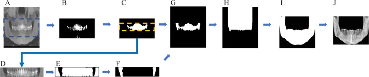
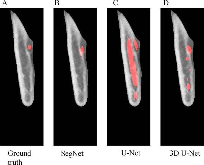
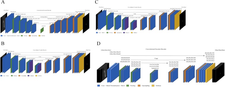
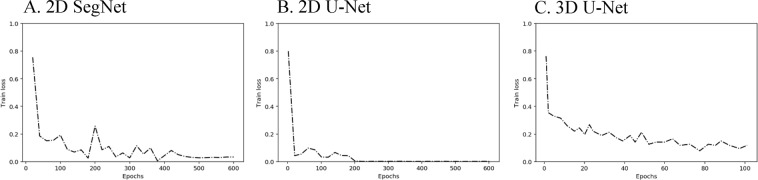
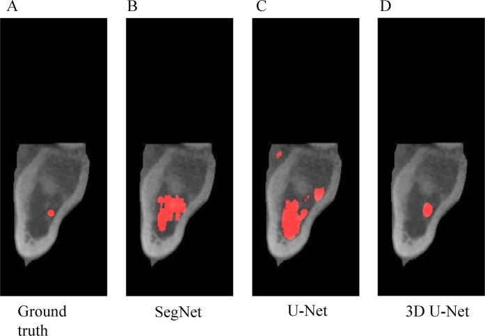
The practicability of deep learning techniques has been demonstrated by their successful implementation in varied fields, including diagnostic imaging for clinicians. In accordance with the increasing demands in the healthcare industry, techniques for automatic prediction and detection are being widely researched. Particularly in dentistry, for various reasons, automated mandibular canal detection has become highly desirable. The positioning of the inferior alveolar nerve (IAN), which is one of the major structures in the mandible, is crucial to prevent nerve injury during surgical procedures. However, automatic segmentation using Cone beam computed tomography (CBCT) poses certain difficulties, such as the complex appearance of the human skull, limited number of datasets, unclear edges, and noisy images. Using work-in-progress automation software, experiments were conducted with models based on 2D SegNet, 2D and 3D U-Nets as preliminary research for a dental segmentation automation tool. The 2D U-Net with adjacent images demonstrates higher global accuracy of 0.82 than naïve U-Net variants. The 2D SegNet showed the second highest global accuracy of 0.96, and the 3D U-Net showed the best global accuracy of 0.99. The automated canal detection system through deep learning will contribute significantly to efficient treatment planning and to reducing patients' discomfort by a dentist. This study will be a preliminary report and an opportunity to explore the application of deep learning to other dental fields.

摘要

深度学习技术的实用性已经在各种领域得到了证明,包括临床医生的诊断成像。根据医疗保健行业的不断增长的需求,自动预测和检测技术正在被广泛研究。特别是在牙科领域,由于各种原因,自动检测下颌管的需求变得非常迫切。定位下颌神经(IAN),这是下颌骨中的主要结构之一,对于防止手术过程中的神经损伤至关重要。然而,使用锥形束计算机断层扫描(CBCT)进行自动分割存在一定的困难,例如人类颅骨的复杂外观、数据集数量有限、边缘不清晰和图像噪声等。使用正在进行中的自动化软件,基于 2D SegNet、2D 和 3D U-Net 的模型进行了实验,作为牙科分割自动化工具的初步研究。使用相邻图像的 2D U-Net 比原始 U-Net 变体具有更高的全局准确性,为 0.82。2D SegNet 显示第二高的全局准确性,为 0.96,3D U-Net 显示最佳的全局准确性,为 0.99。通过深度学习的自动管检测系统将为有效的治疗计划和减少患者的不适做出重大贡献。本研究将是一个初步报告,并为探索深度学习在其他牙科领域的应用提供机会。

https://cdn.ncbi.nlm.nih.gov/pmc/blobs/0419/7109125/638bac6f174e/41598_2020_62586_Fig5_HTML.jpg
https://cdn.ncbi.nlm.nih.gov/pmc/blobs/0419/7109125/bc3b820c93b2/41598_2020_62586_Fig1_HTML.jpg
https://cdn.ncbi.nlm.nih.gov/pmc/blobs/0419/7109125/bbf19c6bdc9a/41598_2020_62586_Fig2_HTML.jpg
https://cdn.ncbi.nlm.nih.gov/pmc/blobs/0419/7109125/4ca215c5c21d/41598_2020_62586_Fig3_HTML.jpg
https://cdn.ncbi.nlm.nih.gov/pmc/blobs/0419/7109125/12a288c22898/41598_2020_62586_Fig4_HTML.jpg
https://cdn.ncbi.nlm.nih.gov/pmc/blobs/0419/7109125/638bac6f174e/41598_2020_62586_Fig5_HTML.jpg

相似文献

1
Automatic mandibular canal detection using a deep convolutional neural network.使用深度卷积神经网络进行自动下颌管检测。
Sci Rep. 2020 Mar 31;10(1):5711. doi: 10.1038/s41598-020-62586-8.
2
Towards clinically applicable automated mandibular canal segmentation on CBCT.实现 CBCT 下颌管临床应用的自动分割
J Dent. 2024 May;144:104931. doi: 10.1016/j.jdent.2024.104931. Epub 2024 Mar 6.
3
Risk assessment of inferior alveolar nerve injury for immediate implant placement in the posterior mandible: a virtual implant placement study.下颌后牙区即刻种植时下牙槽神经损伤的风险评估:一项虚拟种植体植入研究
J Dent. 2014 Mar;42(3):263-70. doi: 10.1016/j.jdent.2013.12.014. Epub 2014 Jan 3.
4
Deep Learning Method for Mandibular Canal Segmentation in Dental Cone Beam Computed Tomography Volumes.基于口腔锥形束 CT 容积数据的下颌管分割的深度学习方法。
Sci Rep. 2020 Apr 3;10(1):5842. doi: 10.1038/s41598-020-62321-3.
5
Relationship between the position of the mental foramen and the anterior loop of the inferior alveolar nerve as determined by cone beam computed tomography combined with mimics.锥形束计算机断层扫描结合Mimics软件测定颏孔位置与下牙槽神经前袢的关系
J Comput Assist Tomogr. 2015 Jan-Feb;39(1):86-93. doi: 10.1097/RCT.0000000000000166.
6
Layered deep learning for automatic mandibular segmentation in cone-beam computed tomography.基于分层深度学习的锥形束计算机断层扫描下颌骨自动分割。
J Dent. 2021 Nov;114:103786. doi: 10.1016/j.jdent.2021.103786. Epub 2021 Aug 20.
7
Automatic detection of inferior alveolar nerve canal from cone-beam computed tomography images for dental surgery planning.
Stud Health Technol Inform. 2014;196:61-5.
8
[A PhD completed. Pre-surgical assessment of the mandibular body and canal using CBCT imaging.].
Ned Tijdschr Tandheelkd. 2018 Jun;125(6):341-344. doi: 10.5177/ntvt.2018.06.18127.
9
Alveolar nerve unfolding technique for synoptic analysis: visualization and quantification of inferior alveolar nerve tracings in three-dimensional cone-beam computed tomography.用于概要分析的牙槽神经展开技术:三维锥形束计算机断层扫描中下牙槽神经走行的可视化与量化
J Craniofac Surg. 2013 Jul;24(4):e374-7. doi: 10.1097/SCS.0b013e3182903013.
10
Position of the impacted third molar in relation to the mandibular canal. Diagnostic accuracy of cone beam computed tomography compared with panoramic radiography.阻生第三磨牙相对于下颌管的位置。锥束计算机断层扫描与全景X线摄影的诊断准确性比较。
Int J Oral Maxillofac Surg. 2009 Sep;38(9):964-71. doi: 10.1016/j.ijom.2009.06.007. Epub 2009 Jul 28.

引用本文的文献

1
Assessing the spatial relationship between mandibular third molars and the inferior alveolar canal using a deep learning-based approach: a proof-of-concept study.使用基于深度学习的方法评估下颌第三磨牙与下牙槽神经管之间的空间关系:一项概念验证研究。
BMC Oral Health. 2025 Aug 6;25(1):1297. doi: 10.1186/s12903-025-06539-5.
2
Clinical application of a deep learning system for automatic mandibular alveolar bone quantity assessment and suggested treatment options using CBCT cross-sections.深度学习系统在利用锥形束计算机断层扫描(CBCT)横断面自动评估下颌牙槽骨量及建议治疗方案中的临床应用
Medicine (Baltimore). 2025 Jul 25;104(30):e43257. doi: 10.1097/MD.0000000000043257.
3

本文引用的文献

1
An overview of deep learning in the field of dentistry.牙科领域深度学习概述。
Imaging Sci Dent. 2019 Mar;49(1):1-7. doi: 10.5624/isd.2019.49.1.1. Epub 2019 Mar 25.
2
Anatomical position of the mandibular canal in relation to the buccal cortical bone: relevance to sagittal split osteotomy.下颌管相对于颊侧皮质骨的解剖位置:与矢状劈开截骨术的相关性。
J Korean Assoc Oral Maxillofac Surg. 2018 Aug;44(4):167-173. doi: 10.5125/jkaoms.2018.44.4.167. Epub 2018 Aug 29.
3
Computer-aided diagnosis of lung nodule classification between benign nodule, primary lung cancer, and metastatic lung cancer at different image size using deep convolutional neural network with transfer learning.
Automatic Segmentation of the Infraorbital Canal in CBCT Images: Anatomical Structure Recognition Using Artificial Intelligence.
锥形束计算机断层扫描(CBCT)图像中眶下管的自动分割:基于人工智能的解剖结构识别
Diagnostics (Basel). 2025 Jul 4;15(13):1713. doi: 10.3390/diagnostics15131713.
4
Assessment of the Diagnostic Accuracy of Artificial Intelligence Software in Identifying Common Periodontal and Restorative Dental Conditions (Marginal Bone Loss, Periapical Lesion, Crown, Restoration, Dental Caries) in Intraoral Periapical Radiographs.评估人工智能软件在识别口腔根尖片上常见牙周和修复性牙科病症(边缘骨丧失、根尖周病变、牙冠、修复体、龋齿)方面的诊断准确性。
Diagnostics (Basel). 2025 Jun 4;15(11):1432. doi: 10.3390/diagnostics15111432.
5
AI-powered segmentation of bifid mandibular canals using CBCT.使用锥形束计算机断层扫描(CBCT)对下颌骨双管进行人工智能驱动的分割。
BMC Oral Health. 2025 Jun 4;25(1):907. doi: 10.1186/s12903-025-06311-9.
6
Deploying a novel deep learning framework for segmentation of specific anatomical structures on cone-beam CT.部署一种用于在锥束CT上分割特定解剖结构的新型深度学习框架。
Oral Radiol. 2025 May 30. doi: 10.1007/s11282-025-00831-4.
7
Accuracy of artificial intelligence-based segmentation in maxillofacial structures: a systematic review.基于人工智能的颌面结构分割的准确性:一项系统综述。
BMC Oral Health. 2025 Mar 7;25(1):350. doi: 10.1186/s12903-025-05730-y.
8
Segmentation of the nasopalatine canal and detection of canal furcation status with artificial intelligence on cone-beam computed tomography images.锥形束计算机断层扫描图像上利用人工智能对鼻腭管进行分割并检测管分叉状态
Oral Radiol. 2025 Feb 28. doi: 10.1007/s11282-025-00812-7.
9
Automated Age Estimation from OPG Images and Patient Records Using Deep Feature Extraction and Modified Genetic-Random Forest.使用深度特征提取和改进的遗传随机森林从全景曲面断层(OPG)图像和患者记录中进行自动年龄估计
Diagnostics (Basel). 2025 Jan 29;15(3):314. doi: 10.3390/diagnostics15030314.
10
Artificial intelligence in dentistry and dental biomaterials.牙科及牙科生物材料中的人工智能
Front Dent Med. 2024 Dec 23;5:1525505. doi: 10.3389/fdmed.2024.1525505. eCollection 2024.
基于深度卷积神经网络的迁移学习在不同图像大小下对肺结节良恶性、原发性肺癌和转移性肺癌进行计算机辅助诊断。
PLoS One. 2018 Jul 27;13(7):e0200721. doi: 10.1371/journal.pone.0200721. eCollection 2018.
4
3-D Convolutional Encoder-Decoder Network for Low-Dose CT via Transfer Learning From a 2-D Trained Network.基于二维网络训练的迁移学习的用于低剂量 CT 的三维卷积编解码器网络。
IEEE Trans Med Imaging. 2018 Jun;37(6):1522-1534. doi: 10.1109/TMI.2018.2832217.
5
Anatomic study of the position of the mandibular canal and corresponding mandibular third molar on cone-beam computed tomography images.锥形束计算机断层扫描图像上下颌管位置及相应下颌第三磨牙的解剖学研究。
Surg Radiol Anat. 2018 Jun;40(6):609-614. doi: 10.1007/s00276-017-1928-6. Epub 2017 Oct 27.
6
Mandibular Canal Enlargement: Clinical and Radiological Characteristics.下颌管扩大:临床与影像学特征
J Clin Imaging Sci. 2017 Jul 13;7:28. doi: 10.4103/jcis.JCIS_28_17. eCollection 2017.
7
SegNet: A Deep Convolutional Encoder-Decoder Architecture for Image Segmentation.SegNet:一种用于图像分割的深度卷积编解码器架构。
IEEE Trans Pattern Anal Mach Intell. 2017 Dec;39(12):2481-2495. doi: 10.1109/TPAMI.2016.2644615. Epub 2017 Jan 2.
8
Automatic segmentation of mandibular canal in cone beam CT images using conditional statistical shape model and fast marching.基于条件统计形状模型和快速行进法的锥形束 CT 图像下颌管自动分割
Int J Comput Assist Radiol Surg. 2017 Apr;12(4):581-593. doi: 10.1007/s11548-016-1484-2. Epub 2016 Sep 21.
9
Effective dose of cone beam CT (CBCT) of the facial skeleton: a systematic review.面部骨骼锥形束CT(CBCT)的有效剂量:一项系统评价。
Br J Radiol. 2015 Jan;88(1045):20140658. doi: 10.1259/bjr.20140658.
10
Radiographic evaluation of the course and visibility of the mandibular canal.下颌管走行及显影情况的影像学评估。
Imaging Sci Dent. 2014 Dec;44(4):273-8. doi: 10.5624/isd.2014.44.4.273. Epub 2014 Nov 25.