Department of Brain Sciences, Faculty of Medicine, Imperial College, London, UK.
Merck Healthcare KGaA, Darmstadt, Germany.
Brain Pathol. 2020 Jul;30(4):779-793. doi: 10.1111/bpa.12841. Epub 2020 Apr 26.
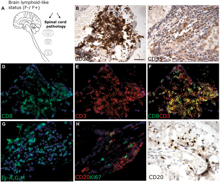
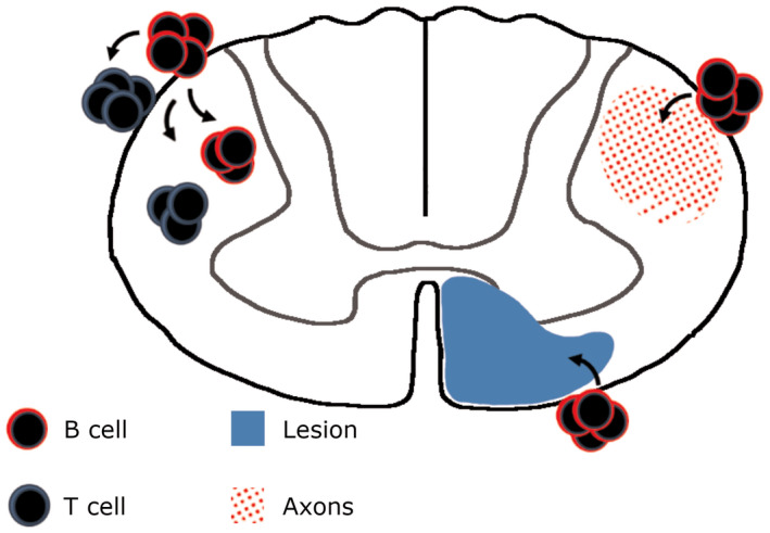
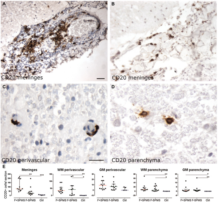
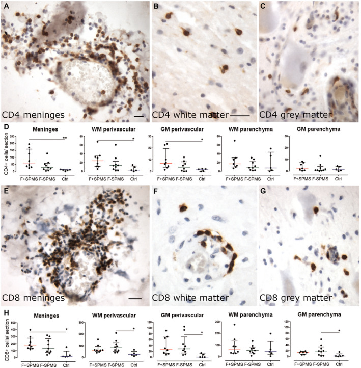
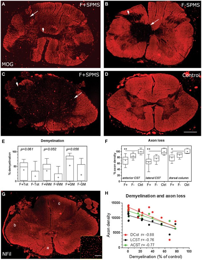
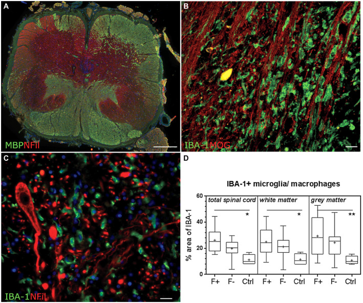
Increased inflammation in the cerebral meninges is associated with extensive subpial cortical grey matter pathology in the forebrain and a more severe disease course in a substantial proportion of secondary progressive multiple sclerosis (SPMS) cases. It is not known whether this relationship extends to spinal cord pathology. We assessed the contribution of meningeal and parenchymal immune infiltrates to spinal cord pathology in SPMS cases characterized in the presence (F+) or absence (F-) of lymphoid-like structures in the forebrain meninges. Transverse cryosections of cervical, thoracic and lumbar cord of 22 SPMS and five control cases were analyzed for CD20+ B cells, CD4+ and CD8+ T cells, microglia/macrophages (IBA-1+), demyelination (myelin oligodendrocyte glycoprotein+) and axon density (neurofilament-H+). Lymphoid-like structures containing follicular dendritic cell networks and dividing B cells were seen in the spinal meninges of 3 out of 11 F+ SPMS cases. CD4+ and CD20+ cell counts were increased in F+ SPMS compared to F- SPMS and controls, whilst axon loss was greatest in motor and sensory tracts of the F+ SPMS cases (P < 0.01). The density of CD20+ B cells of the spinal leptomeninges correlated with CD4+ T cells and total B and T cells of the meninges; with the density of white matter perivascular CD20+ and CD4+ lymphocytes (P < 0.05); with white matter lesion area (P < 0.05); and the extent of axon loss (P < 0.05) in F+ SPMS cases only. We show that the presence of lymphoid-like structures in the forebrain is associated with a profound spinal cord pathology and local B cell rich meningeal inflammation associates with the extent of cord pathology. Our work supports a principal role for B cells in sustaining inflammation and tissue injury throughout the CNS in the progressive disease stage.
大脑脑膜中的炎症与前脑广泛的皮质下灰质病理有关,并且在相当一部分继发进展性多发性硬化症(SPMS)病例中,疾病进程更为严重。目前尚不清楚这种关系是否会扩展到脊髓病理。我们评估了脑膜和实质免疫浸润物对 SPMS 病例脊髓病理的贡献,这些病例在前脑脑膜中存在(F+)或不存在(F-)淋巴样结构。对 22 例 SPMS 和 5 例对照病例的颈、胸和腰段脊髓的横断冷冻切片进行了 CD20+B 细胞、CD4+和 CD8+T 细胞、小胶质细胞/巨噬细胞(IBA-1+)、脱髓鞘(髓鞘少突胶质细胞糖蛋白+)和轴突密度(神经丝-H+)分析。在 11 例 F+SPMS 病例中的 3 例中,脊髓脑膜中可见包含滤泡树突状细胞网络和分裂 B 细胞的淋巴样结构。与 F- SPMS 和对照组相比,F+ SPMS 中的 CD4+和 CD20+细胞计数增加,而 F+ SPMS 病例的运动和感觉束中轴突丢失最大(P<0.01)。脊髓软脑膜的 CD20+B 细胞密度与脑膜中的 CD4+T 细胞和总 B 细胞和 T 细胞、与白质血管周围 CD20+和 CD4+淋巴细胞密度(P<0.05)、与白质病变面积(P<0.05)和轴突丢失程度(P<0.05)相关仅在 F+ SPMS 病例中。我们表明,在前脑存在淋巴样结构与严重的脊髓病理有关,局部富含 B 细胞的脑膜炎症与脊髓病理的程度有关。我们的工作支持 B 细胞在进展性疾病阶段维持整个中枢神经系统炎症和组织损伤中的主要作用。