School of Biomedical Engineering, The University of British Columbia, Vancouver, B.C., Canada.
Michael Cuccione Childhood Cancer Research Program, BC Children's Hospital Research Institute, Vancouver, B.C., Canada.
PLoS One. 2020 Apr 3;15(4):e0230966. doi: 10.1371/journal.pone.0230966. eCollection 2020.
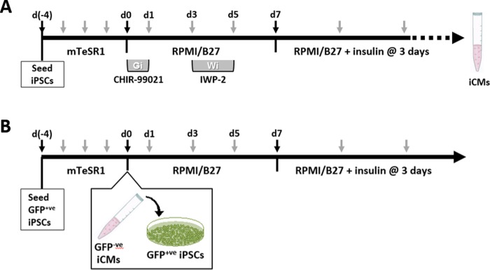
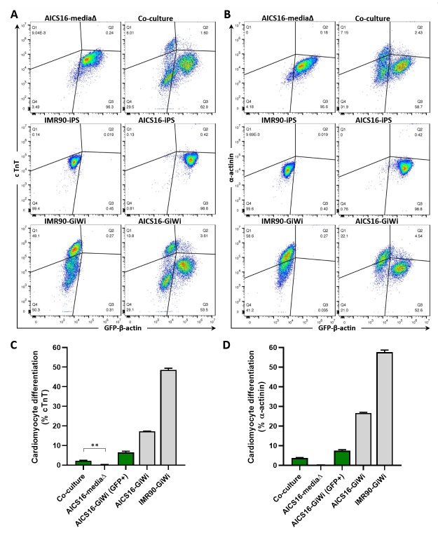
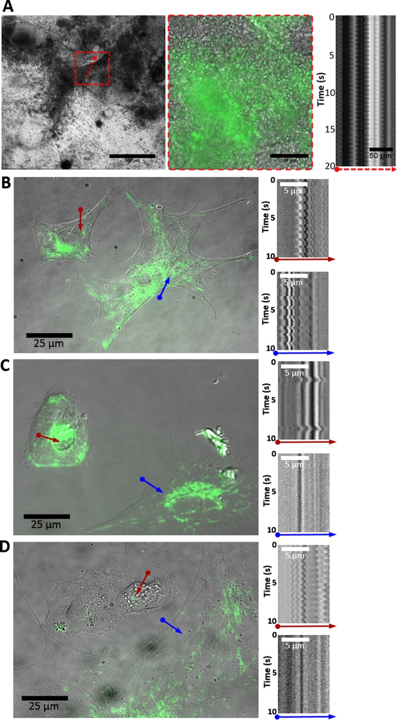
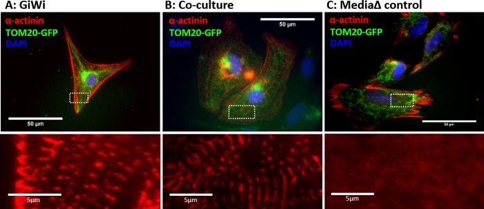
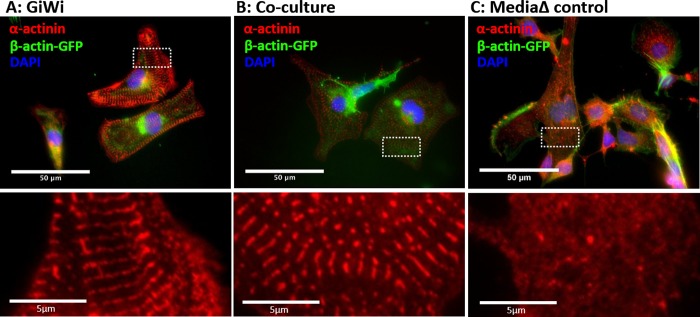
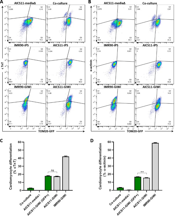
Various types of stem cells and non-stem cells have been shown to differentiate or transdifferentiate into cardiomyocytes by way of co-culture with appropriate inducer cells. However, there is a limited demonstration of a co-culture induction system utilizing stem cell-derived cardiomyocytes as a stimulatory source for cardiac reprogramming (of stem cells or otherwise). In this study, we utilized an inductive co-culture method to show that previously differentiated induced pluripotent stem (iPS) cell-derived cardiomyocytes (iCMs), when co-cultivated with iPS cells, constituted a sufficient stimulatory system to induce cardiac differentiation. To enable tracking of both cell populations, we utilized GFP-labeled iPS cells and non-labeled iCMs pre-differentiated using inhibitors of GSK and Wnt signaling. Successful differentiation was assessed by the exhibition of spontaneous self-contractions, structural organization of α-actinin labeled sarcomeres, and expression of cardiac specific markers cTnT and α-actinin. We found that iCM-iPS cell-cell contact was essential for inductive differentiation, and this required overlaying already adherent iPS cells with iCMs. Importantly, this process was achieved without the exogenous addition of pathway inhibitors and morphogens, suggesting that 'older' iCMs serve as an adequate stimulatory source capable of recapitulating the necessary culture environment for cardiac differentiation.
各种类型的干细胞和非干细胞已经通过与适当的诱导细胞共培养显示出分化或转分化为心肌细胞。然而,利用干细胞来源的心肌细胞作为心脏重编程(干细胞或其他)的刺激源的共培养诱导系统的证明是有限的。在这项研究中,我们利用诱导共培养方法表明,先前分化的诱导多能干细胞(iPS)细胞衍生的心肌细胞(iCMs),与 iPS 细胞共培养时,构成了诱导心脏分化的充分刺激系统。为了能够同时追踪两个细胞群体,我们利用 GFP 标记的 iPS 细胞和使用 GSK 和 Wnt 信号通路抑制剂预先分化的非标记 iCMs。通过自发的自我收缩、标记有肌动蛋白的肌节的结构组织以及心脏特异性标志物 cTnT 和肌动蛋白的表达来评估成功的分化。我们发现 iCM-iPS 细胞间的细胞接触对于诱导分化是必不可少的,这需要用 iCMs 覆盖已经附着的 iPS 细胞。重要的是,这个过程是在没有添加外源途径抑制剂和形态发生素的情况下实现的,这表明“较老”的 iCMs 可以作为一种足够的刺激源,能够再现心脏分化所需的必要培养环境。