Université de Nantes, Inserm, CRCINA, F-44000, Nantes, France.
LabEx IGO, Université de Nantes, Nantes, France.
Sci Rep. 2020 Apr 3;10(1):5900. doi: 10.1038/s41598-020-62664-x.
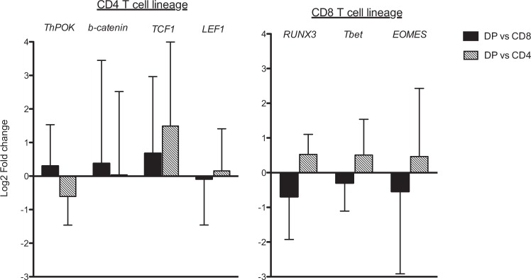
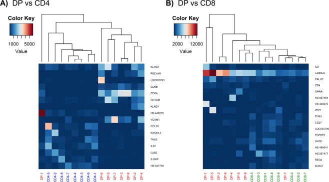
Peripheral CD4CD8 double positive (DP) T cells are a phenotypically and functionally heterogeneous population depending on their origin and pathologic context. We previously identified among tumour infiltrating lymphocytes in melanoma, a tumour-reactive MHC class-I restricted CD4CD8 DP αβ T-cell subpopulation with CD4-like function. In this study, we used an in-depth comparative transriptomic analysis of intra-melanoma DP T cells and CD4 and CD8 single positive (SP) T cells, to better comprehend the origin of this DP phenotype, and define the transcriptomic signature of activated DP T cells. We observed that intra-melanoma DP T cells were transcriptome-wise closer to their CD8 SP T-cell counterparts in terms of number of genes differentially expressed (97 in common with CD8 SP T cells and 15 with CD4 SP T cells) but presented hallmarks of a transition to a CD4-like functional profile (CD40LG) with a decreased cytotoxic signature (KLRC1) in favour of an increased cytokine-receptor interaction signature (IL4, IL24, IL17A…). This unleashed CD4-like program could be the results of the observed unbalanced expression of the THPOK/Runx3 transcription factors in DP T cells. Overall, this study allow us to speculate that intra-melanoma DP T cells arise from CD8 SP T cells being reprogrammed to a helper function.
外周血 CD4CD8 双阳性 (DP) T 细胞是一群表型和功能上具有异质性的细胞群体,具体取决于其起源和病理背景。我们之前在黑色素瘤浸润的淋巴细胞中发现了一种肿瘤反应性 MHC Ⅰ类受限的 CD4CD8 DPαβ T 细胞亚群,具有 CD4 样功能。在这项研究中,我们使用了深入的比较转录组学分析,研究了黑色素瘤内 DP T 细胞和 CD4 和 CD8 单阳性 (SP) T 细胞,以更好地理解这种 DP 表型的起源,并定义激活的 DP T 细胞的转录组特征。我们观察到,就差异表达基因的数量而言,黑色素瘤内 DP T 细胞在转录组上更接近其 CD8 SP T 细胞的对应物(与 CD8 SP T 细胞共有 97 个,与 CD4 SP T 细胞共有 15 个),但表现出向 CD4 样功能表型(CD40LG)转变的特征,其细胞毒性特征(KLRC1)降低,而细胞因子受体相互作用特征(IL4、IL24、IL17A……)增加。这种被释放的 CD4 样程序可能是 DP T 细胞中观察到的 THPOK/Runx3 转录因子表达不平衡的结果。总的来说,这项研究使我们推测黑色素瘤内 DP T 细胞可能是由 CD8 SP T 细胞重新编程为辅助功能而来。