Department of Psychiatry and Behavioral Medicine, Medical College of Wisconsin (GC, SAC, SJL, JSG), Milwaukee, WI; Department of Biophysics, Medical College of Wisconsin (GC, BDW, SJL), Milwaukee, WI.
Department of Biophysics, Medical College of Wisconsin (GC, BDW, SJL), Milwaukee, WI.
Am J Geriatr Psychiatry. 2020 Oct;28(10):1089-1101. doi: 10.1016/j.jagp.2020.02.014. Epub 2020 Mar 9.
Acute grief, in an important minority of older adults, can become protracted, intense, and debilitating, leading to the development of complicated grief (CG). However, the neurobiologic mechanisms underlying a maladaptive grief response after an attachment loss are unknown. The current study aimed to examine the amygdala brain network features that cross-sectionally explain the symptom variance and longitudinally relate to grief symptom trajectories after an attachment loss.
Baseline amygdala functional connectivity (Fc) was assessed using a seed-based resting-state functional magnetic resonance imaging method in 35 adults who were within 1-year after death of a loved one and 21 healthy comparison (HC) participants. Magnetic resonance imaging scans were obtained at baseline, and clinical assessments, including the inventory of complicated grief (ICG) were completed at weeks 0, 8, 16, and 26 (endpoint).
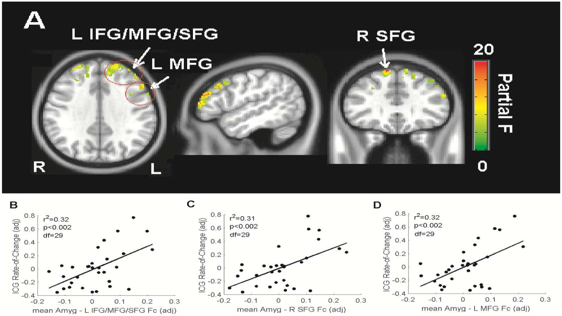
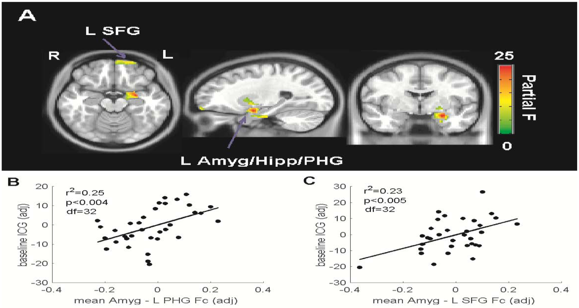
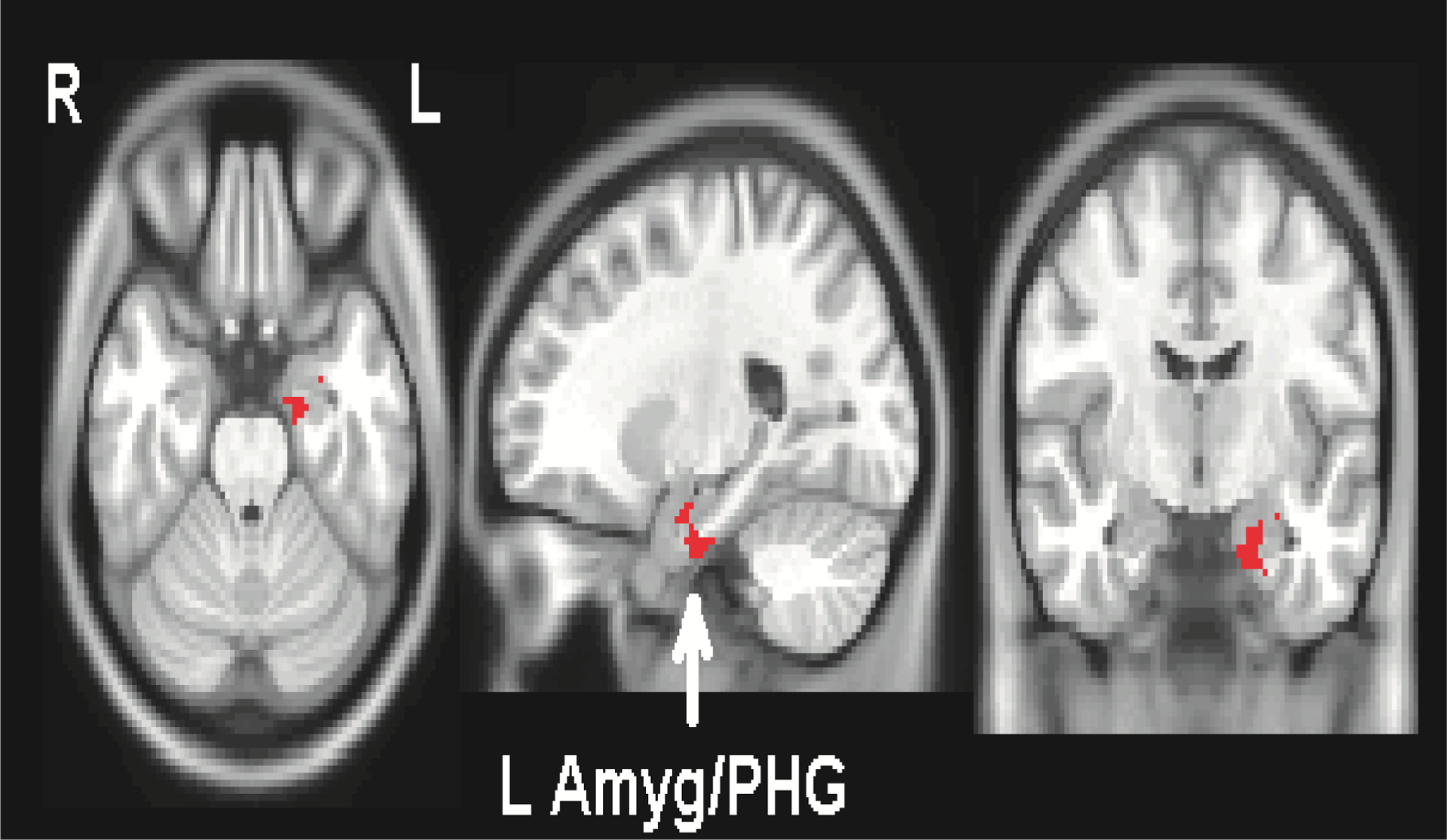
Relative to HC participants, grief participants showed increased amygdala Fc in the posterior default mode (bilateral medial temporal lobes and left precuneus) and thalamus. Amygdala Fc in the default mode and ventral affective regions positively correlated with ICG scores at baseline. Furthermore, increased baseline amygdala functional connections with the dorsal frontal executive control and salience network regions correlated with worsening ICG scores over time. These longitudinal findings persisted after controlling for covariates, including baseline depressive and anxiety symptoms.
These results provide novel preliminary evidence suggesting amygdala-based brain network measures to cross-sectionally explain symptom variance and longitudinally correlate with grief symptom trajectories in grievers. Amygdala brain network function measures may have the potential to serve as biomarkers of CG.
在少数老年人中,急性悲伤可能会变得持久、强烈和衰弱,导致复杂悲伤(CG)的发展。然而,在失去依恋后,适应不良的悲伤反应的神经生物学机制尚不清楚。本研究旨在探讨杏仁核脑网络特征,这些特征可横向解释症状变异,并与失去依恋后悲伤症状轨迹纵向相关。
使用基于种子的静息态功能磁共振成像方法,在 35 名失去亲人后 1 年内的成年人和 21 名健康对照(HC)参与者中评估基线杏仁核功能连接(Fc)。在基线时进行磁共振成像扫描,并在第 0、8、16 和 26 周(终点)进行临床评估,包括复杂悲伤量表(ICG)。
与 HC 参与者相比,悲伤参与者在默认模式的后杏仁核(双侧内侧颞叶和左楔前叶)和丘脑中表现出增加的杏仁核 Fc。默认模式和腹侧情感区域的杏仁核 Fc 与基线时的 ICG 评分呈正相关。此外,基线时杏仁核与背侧额叶执行控制和突显网络区域的功能连接增加与随时间推移 ICG 评分的恶化相关。这些纵向发现在控制了基线抑郁和焦虑症状等协变量后仍然存在。
这些结果提供了新的初步证据,表明基于杏仁核的脑网络测量可横向解释症状变异,并与悲伤者的悲伤症状轨迹纵向相关。杏仁核脑网络功能测量可能有潜力成为 CG 的生物标志物。