Fan Zhongjun, Wang Huanli, Pan Jiahao, Yu Shupei, Xia Wenlong
College of Marine and Biological Engineering, Yancheng Teachers' University, Yancheng 224002, China.
J Vet Res. 2020 Jan 31;64(1):33-38. doi: 10.2478/jvetres-2020-0009. eCollection 2020 Mar.
Marek's disease virus (MDV) can cause malignant T-cell lymphomas and immunosuppression in chickens. Macrophage migration inhibitory factor (MIF) not only plays a critical role in inhibiting T-cell responses, but also contributes to multiple aspects of tumour progression. The aim of this study was to reveal the potential role of MIF in the pathogenesis of MDV infection.
MIF gene expression levels were measured by using real-time PCR. Expression was assayed at different times in chicken embryo fibroblast (CEF) cells and tissue samples of SPF chickens infected with different MDV strains and fold change was calculated by the 2 method.
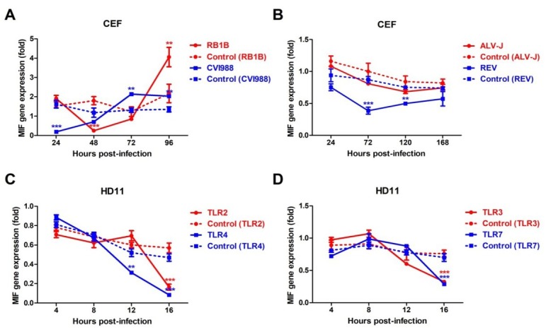
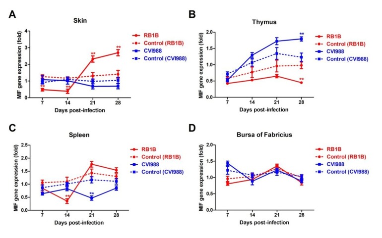
The expression of MIF was significantly downregulated (p < 0.05 and FC > 2) in CEF cells infected with the very virulent MDV RB1B strain at 48 h post infection (hpi) and in the skin and spleen at 14 days post infection (dpi). The reduction of MIF expression was also found in CEF cells infected by reticuloendotheliosis virus (REV), avian leukosis virus subgroup J (ALV-J), and MDV vaccine strain CVI988 or in HD11 cells stimulated with TLR2, 3, 4, and 7 ligands. Interestingly, MIF expression decreased continuously from 7 to 28 dpi in the thymus after RB1B virus infection while it increased after CVI988 virus infection. Upregulated expression of MIF was found in CEF infected with RB1B at 96 hpi and in the spleen and skin at 21 and 28 dpi.
The present study revealed the different expression pattern of MIF in response to MDV infection and indicated that MIF level may be associated with MDV pathogenesis.
马立克氏病病毒(MDV)可导致鸡发生恶性T细胞淋巴瘤并引起免疫抑制。巨噬细胞移动抑制因子(MIF)不仅在抑制T细胞反应中起关键作用,还在肿瘤进展的多个方面发挥作用。本研究旨在揭示MIF在MDV感染发病机制中的潜在作用。
采用实时PCR检测MIF基因表达水平。在感染不同MDV毒株的鸡胚成纤维细胞(CEF)和SPF鸡组织样本的不同时间点检测表达情况,并通过2−ΔΔCT法计算倍数变化。
在感染超强毒MDV RB1B株后48小时(hpi)的CEF细胞中以及感染后14天(dpi)的皮肤和脾脏中,MIF的表达显著下调(p < 0.05且FC > 2)。在感染网状内皮组织增生症病毒(REV)、禽白血病病毒J亚群(ALV-J)以及MDV疫苗株CVI988的CEF细胞中,或在用TLR2、3、4和7配体刺激的HD11细胞中,也发现了MIF表达的降低。有趣的是,RB1B病毒感染后,胸腺中MIF表达从7 dpi到28 dpi持续下降,而CVI988病毒感染后则升高。在感染RB1B后96 hpi的CEF细胞以及21和28 dpi的脾脏和皮肤中,发现MIF表达上调。
本研究揭示了MIF在应对MDV感染时的不同表达模式,并表明MIF水平可能与MDV发病机制相关。