Department of Conservative Dentistry, Semmelweis University, Szentkirályi utca 47, Budapest, H-1088, Hungary.
Department of Oral Rehabilitation, Medical University of South Carolina College of Dental Medicine, Charleston, SC, USA.
BMC Oral Health. 2020 Apr 7;20(1):97. doi: 10.1186/s12903-020-01090-x.
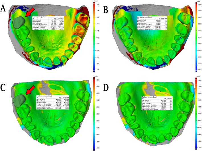
Intraoral scanner (IOS) accuracy is commonly evaluated using full-arch surface comparison, which fails to take into consideration the starting position of the scanning (scan origin). Previously a novel method was developed, which takes into account the scan origin and calculates the deviation of predefined identical points between references and test models. This method may reveal the error caused by stitching individual images during intraoral scan. This study aimed to validate the novel method by comparing the trueness of seven IOSs (Element 1, Element 2, Emerald, Omnicam, Planscan, Trios 3, CS 3600) to a physical impression digitized by laboratory scanner which lacks linear stitching problems.
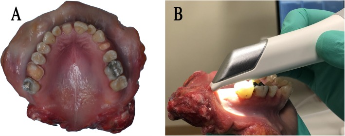
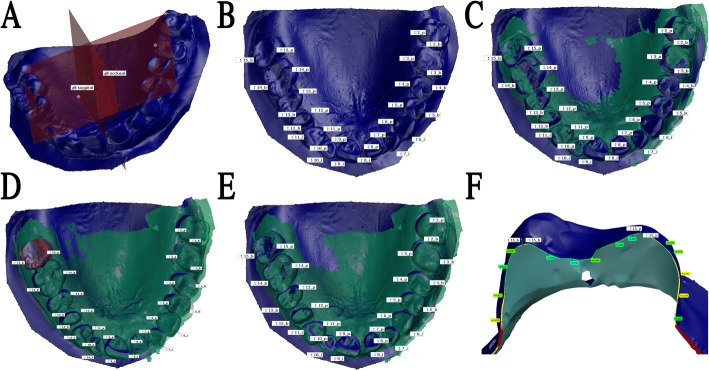
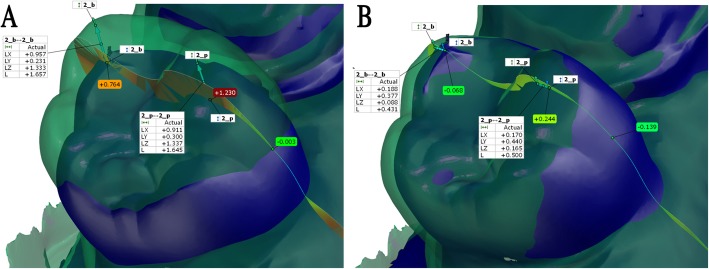
Digital test models of a dentate human cadaver maxilla were made by IOSs and by laboratory scanner after polyvinylsiloxane impression. All scans started on the occlusal surface of the tooth #15 (universal notation, scan origin) and finished at tooth #2. The reference model and test models were superimposed at the scan origin in GOM Inspect software. Deviations were measured between identical points on three different axes, and the complex 3D deviation was calculated. The effect of scanners, tooth, and axis was statistically analyzed by the generalized linear mixed model.
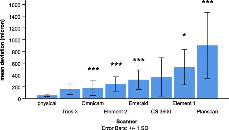
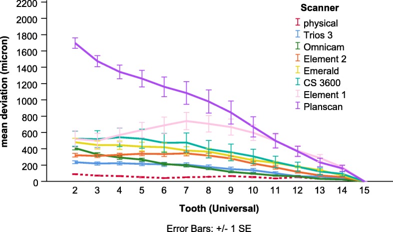
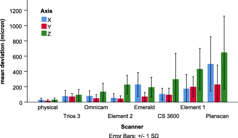
The deviation gradually increased as the distance from scan origin increased for the IOSs but not for the physical impression. The highest deviation occurred mostly at the apico-coronal axis for the IOSs. The mean deviation of the physical impression (53 ± 2 μm) was not significantly different from the Trios 3 (156 ± 8 μm) and CS 3600 (365 ± 29 μm), but it was significantly lower than the values of Element 1 (531 ± 26 μm), Element 2 (246 ± 11 μm), Emerald (317 ± 13 μm), Omnicam (174 ± 11 μm), Planscan (903 ± 49 μm).
The physical impression was superior compared to the IOSs on dentate full-arch of human cadaver. The novel method could reveal the stitching error of IOSs, which may partly be caused by the difficulties in depth measurement.
口腔内扫描仪 (IOS) 的准确性通常通过全牙弓表面比较来评估,但这种方法没有考虑到扫描的起始位置(扫描原点)。先前开发了一种新方法,该方法考虑了扫描原点,并计算了参考模型和测试模型之间预定义相同点的偏差。这种方法可以揭示在口腔内扫描过程中单个图像拼接引起的误差。本研究旨在通过比较七种 IOS(Element 1、Element 2、Emerald、Omnicam、Planscan、Trios 3、CS 3600)与实验室扫描仪数字化的物理印模的准确性来验证新方法,该物理印模缺乏线性拼接问题。
通过 IOS 和聚硅氧烷印模后的实验室扫描仪对人尸体上颌的有牙颌进行数字测试模型制作。所有扫描均从 #15 牙的咬合面(通用标记,扫描原点)开始,在 #2 牙结束。在 GOM Inspect 软件中,将参考模型和测试模型在扫描原点处进行叠加。在三个不同轴上测量相同点之间的偏差,并计算复杂的 3D 偏差。通过广义线性混合模型对扫描仪、牙齿和轴的影响进行统计学分析。
对于 IOS,随着距离扫描原点的增加,偏差逐渐增加,但对于物理印模则不然。对于 IOS,最大偏差大多发生在根尖冠轴上。物理印模的平均偏差(53±2μm)与 Trios 3(156±8μm)和 CS 3600(365±29μm)无显著差异,但明显低于 Element 1(531±26μm)、Element 2(246±11μm)、Emerald(317±13μm)、Omnicam(174±11μm)、Planscan(903±49μm)的数值。
与 IOS 相比,物理印模在人体尸体的全牙弓上具有优势。新方法可以揭示 IOS 的拼接误差,这可能部分是由于深度测量困难造成的。