Department of Anesthesiology, Qilu Hospital of Shandong University, Ji'nan, 250012, China.
Jiangsu Key Laboratory of Drug Screening, China Pharmaceutical University, Nanjing, 210009, China.
J Neuroinflammation. 2020 Apr 7;17(1):109. doi: 10.1186/s12974-020-01799-0.
Accumulating evidence has highlighted the importance of microglial and astrocyte responses in the pathological development of postoperative cognitive dysfunction (POCD). However, the mechanisms involved are not well understood.
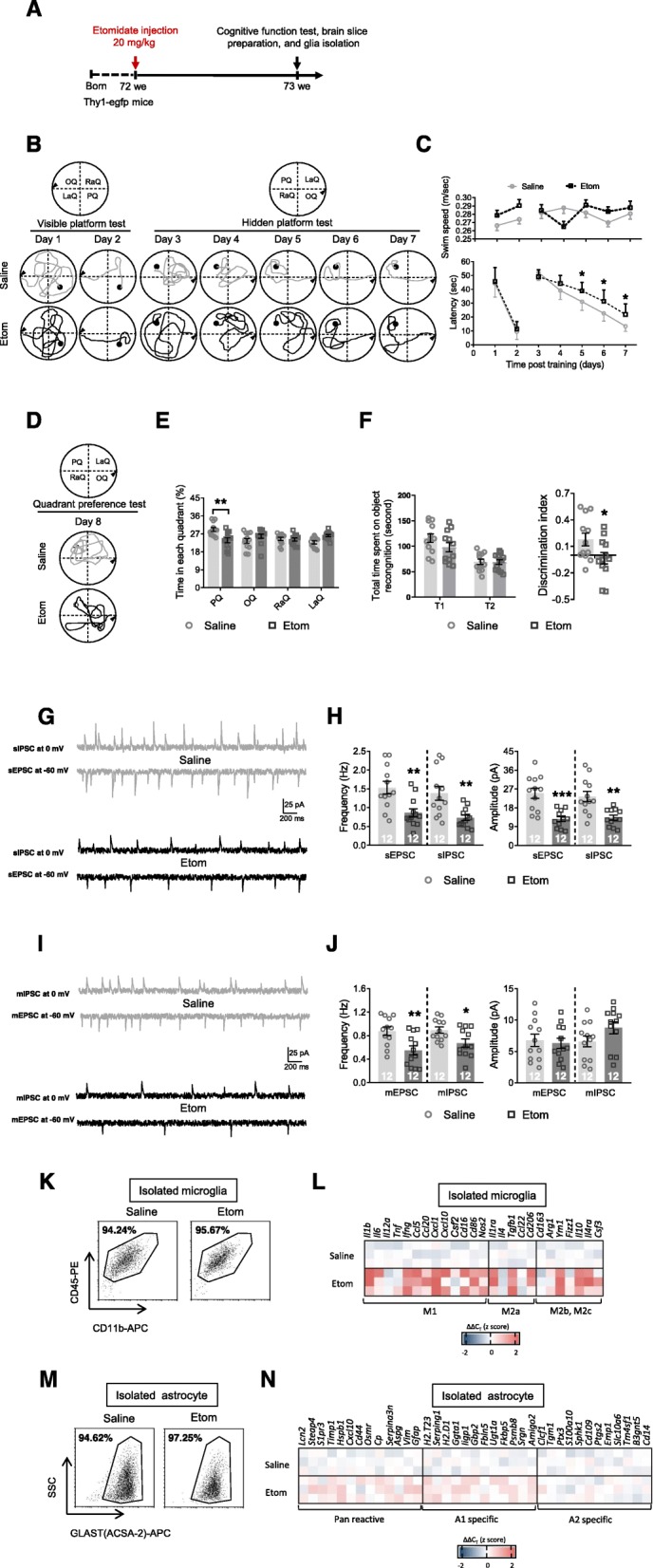
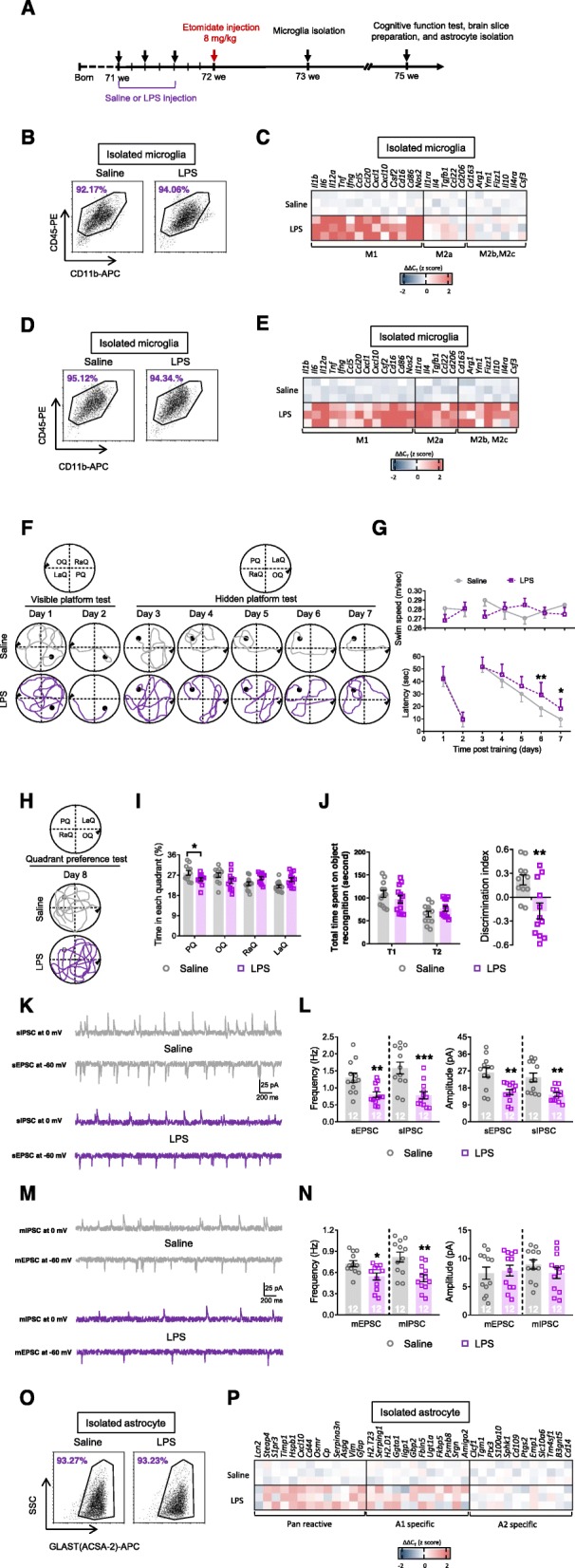
A perioperative neurocognitive disorders (PND) mouse model was generated by administering etomidate, and cognitive function was assessed using the Morris water maze and novel object recognition tests. Excitatory and inhibitory postsynaptic currents were recorded to analyze neuronal activity. In addition, microglia and astrocytes were isolated by magnetic-activated cell sorting, and genes that were activated in these cells were identified using quantitative polymerase chain reaction.
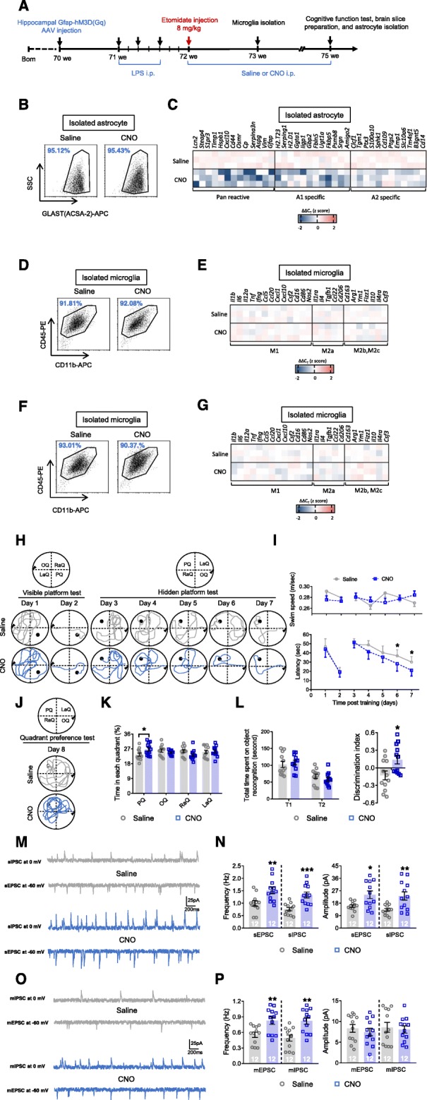
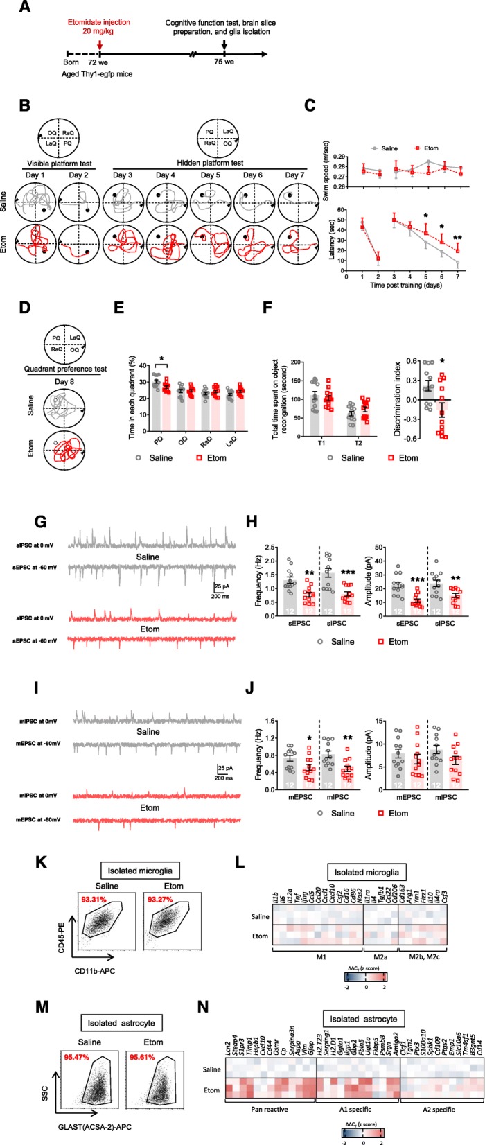
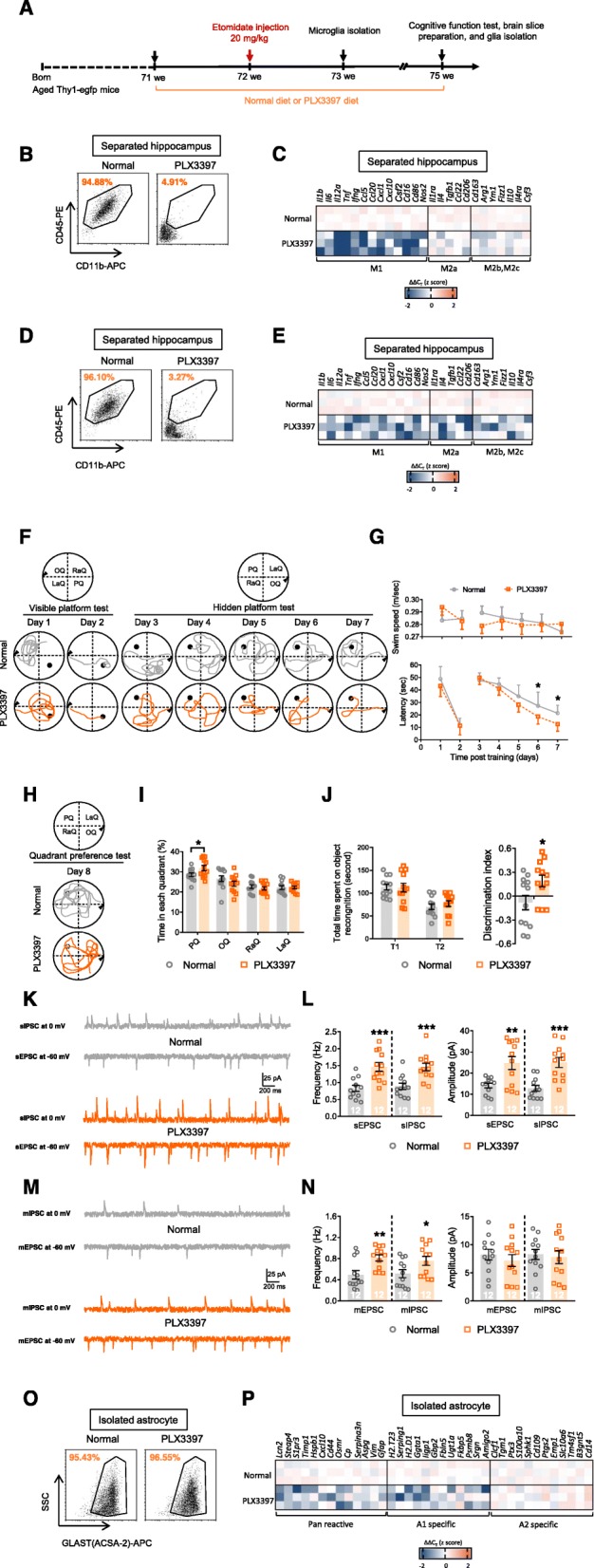
We observed dramatic cognitive impairment at 1 and 3 weeks after etomidate was administered to 18 month-old mice. Microglia and astrocytes isolated from the hippocampus showed significant microglial activation during the early pathological stage (i.e., 1 week after etomidate injection) and an A1-specific astrocyte response during the late pathological stage (i.e., 3 weeks after etomidate injection). Furthermore, when microglia were eliminated before etomidate was injected, the A1-specific astrocyte activation response was significantly reduced, and cognitive function improved. However, when microglia were eliminated after etomidate application, astrocyte activation and cognitive function were not significantly altered. In addition, activating microglia immediately after a sedative dose of etomidate was injected markedly increased A1-specific astrocyte activation and cognitive dysfunction.
A1-specific astrocyte activation is triggered by activated microglia during the initial pathological stage of PND and induces long-term synaptic inhibition and cognitive deficiencies. These results improve our understanding of how PND develops and may suggest therapeutic targets.
越来越多的证据强调了小胶质细胞和星形胶质细胞反应在术后认知功能障碍(POCD)的病理发展中的重要性。然而,其涉及的机制尚不清楚。
通过给予依托咪酯建立围手术期神经认知障碍(PND)小鼠模型,并使用 Morris 水迷宫和新物体识别测试评估认知功能。记录兴奋性和抑制性突触后电流以分析神经元活动。此外,通过磁激活细胞分选分离小胶质细胞和星形胶质细胞,并使用定量聚合酶链反应鉴定这些细胞中激活的基因。
我们观察到在给予 18 月龄小鼠依托咪酯后 1 周和 3 周时,认知功能出现明显障碍。从小鼠海马区分离的小胶质细胞和星形胶质细胞在早期病理阶段(即依托咪酯注射后 1 周)显示出明显的小胶质细胞激活,在晚期病理阶段(即依托咪酯注射后 3 周)显示出 A1 特异性星形胶质细胞反应。此外,在注射依托咪酯前消除小胶质细胞时,A1 特异性星形胶质细胞激活反应显著减少,认知功能得到改善。然而,在依托咪酯应用后消除小胶质细胞时,星形胶质细胞激活和认知功能没有明显改变。此外,在给予镇静剂量依托咪酯后立即激活小胶质细胞,明显增加了 A1 特异性星形胶质细胞激活和认知功能障碍。
在 PND 的初始病理阶段,激活的小胶质细胞触发 A1 特异性星形胶质细胞激活,导致长期突触抑制和认知缺陷。这些结果提高了我们对 PND 发展机制的理解,并可能为治疗靶点提供线索。