• 文献检索
  • 文档翻译
  • 深度研究
  • 学术资讯
  • Suppr Zotero 插件Zotero 插件
  • 邀请有礼
  • 套餐&价格
  • 历史记录
应用&插件
Suppr Zotero 插件Zotero 插件浏览器插件Mac 客户端Windows 客户端微信小程序
定价
高级版会员购买积分包购买API积分包
服务
文献检索文档翻译深度研究API 文档MCP 服务
关于我们
关于 Suppr公司介绍联系我们用户协议隐私条款
关注我们

Suppr 超能文献

核心技术专利:CN118964589B侵权必究
粤ICP备2023148730 号-1Suppr @ 2026

文献检索

告别复杂PubMed语法,用中文像聊天一样搜索,搜遍4000万医学文献。AI智能推荐,让科研检索更轻松。

立即免费搜索

文件翻译

保留排版,准确专业,支持PDF/Word/PPT等文件格式,支持 12+语言互译。

免费翻译文档

深度研究

AI帮你快速写综述,25分钟生成高质量综述,智能提取关键信息,辅助科研写作。

立即免费体验

尼克在大鼠实验性蛛网膜下腔出血后的早期脑损伤中发挥神经保护作用。

Nix Plays a Neuroprotective Role in Early Brain Injury After Experimental Subarachnoid Hemorrhage in Rats.

作者信息

Zhang Juyi, Yuan Guiqiang, Liang Tianyu, Pan Pengjie, Li Xiang, Li Haiying, Shen Haitao, Wang Zhong, Chen Gang

机构信息

Department of Neurosurgery & Brain and Nerve Research Laboratory, The First Affiliated Hospital of Soochow University, Suzhou, China.

出版信息

Front Neurosci. 2020 Mar 24;14:245. doi: 10.3389/fnins.2020.00245. eCollection 2020.

DOI:10.3389/fnins.2020.00245
PMID:32265644
原文链接:https://pmc.ncbi.nlm.nih.gov/articles/PMC7108665/
Abstract

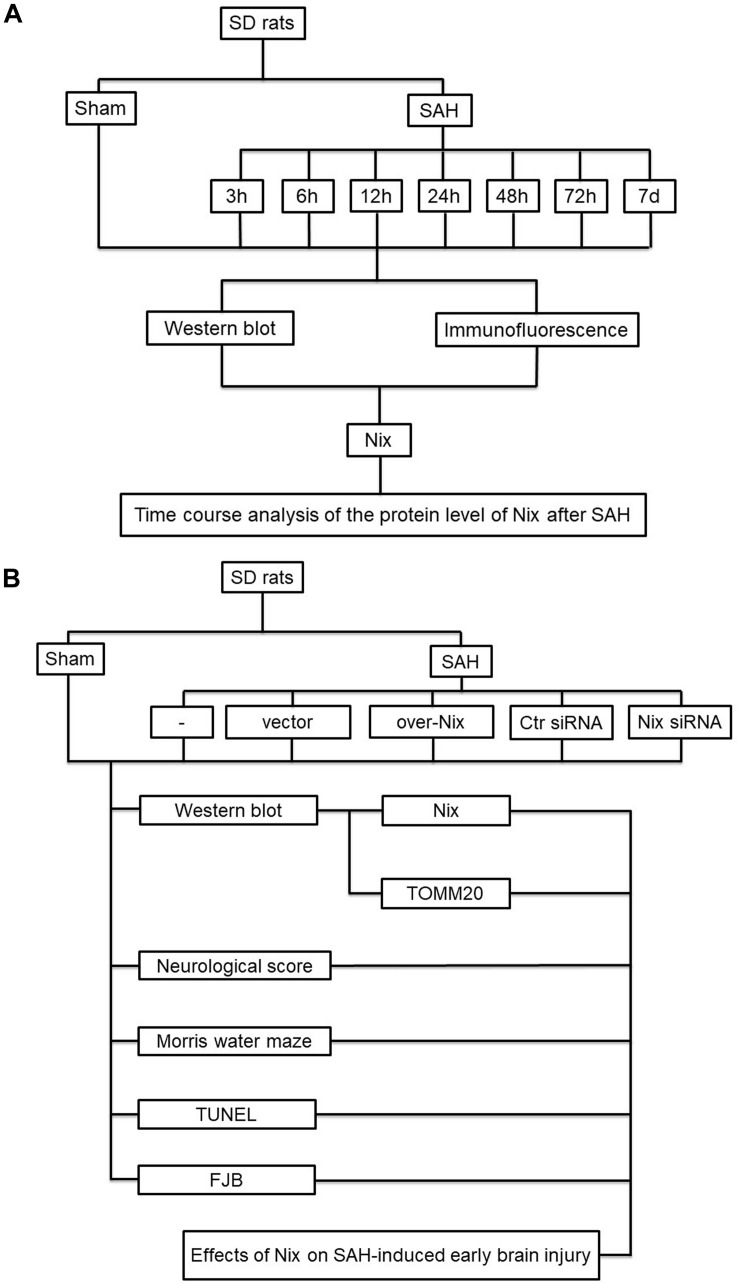
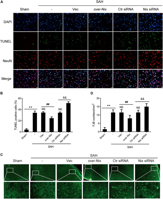
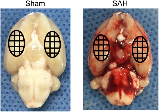
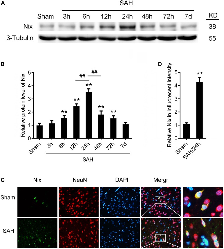
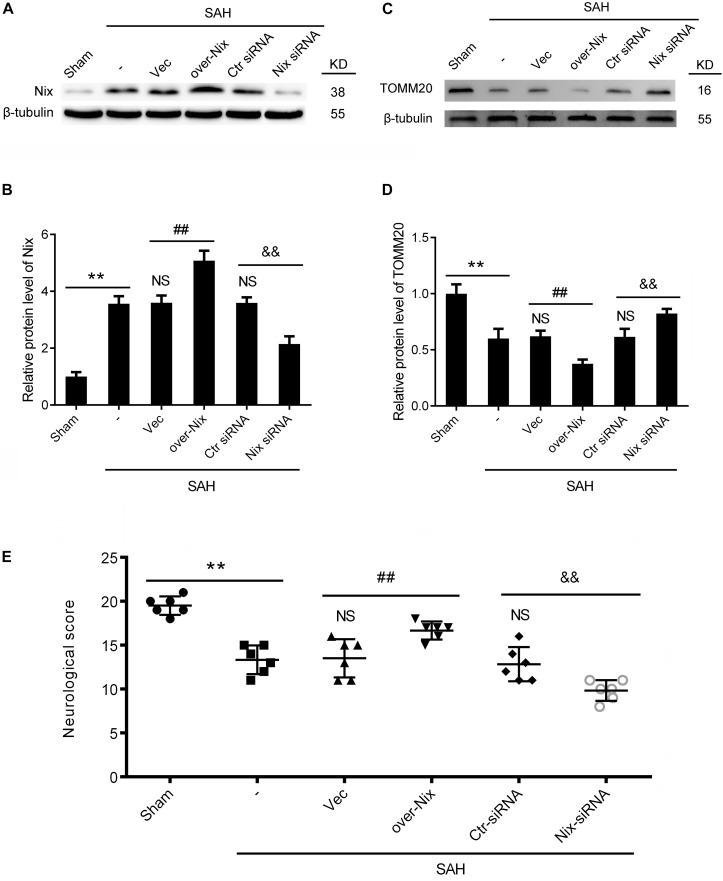
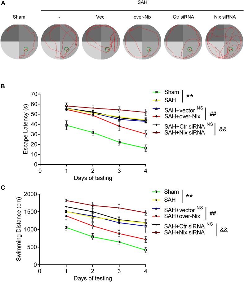
Nix is located in the outer membrane of mitochondria, mediates mitochondrial fission and implicated in many neurological diseases. However, the association between Nix and subarachnoid hemorrhage (SAH) has not previously been reported. Therefore, the present study was designed to evaluate the expression of Nix and its role in early brain injury (EBI) after SAH. Adult male Sprague-Dawley (SD) rats were randomly assigned to various time points for investigation after SAH. A rat model of SAH was induced by injecting 0.3 ml of autologous non-heparinized arterial blood into the prechiasmatic cistern. The expression of Nix was investigated by Western blot and immunohistochemistry. Next, Nix-specific overexpression plasmids and small interfering RNAs (siRNAs) were separately administered. Western blot, neurological scoring, Morris water maze, terminal deoxynucleotidyl transferase-mediated dUTP nick end labeling (TUNEL) staining and fluoro-jade B (FJB) staining were performed to evaluate the role of Nix in EBI following SAH. We found that Nix was expressed in neurons and its expression level in the SAH groups was higher than that in the Sham group, which peaked at 24 h after SAH. Overexpression of Nix following SAH significantly decreased the expression of translocase of outer mitochondrial membrane 20 (TOMM20, a marker of mitochondria), ameliorated neurological/cognitive deficits induced by SAH, and reduced the total number of apoptotic/neurodegenerative cells, whereas siRNA knockdown of Nix yielded opposite effects. Taken together, our findings demonstrated that the expression of Nix is increased in neurons after experimental SAH in rats, and may play a neuroprotective role in EBI following SAH.

摘要

Nix位于线粒体的外膜,介导线粒体分裂,并与许多神经系统疾病有关。然而,此前尚未报道过Nix与蛛网膜下腔出血(SAH)之间的关联。因此,本研究旨在评估Nix的表达及其在SAH后早期脑损伤(EBI)中的作用。成年雄性Sprague-Dawley(SD)大鼠在SAH后被随机分配到不同时间点进行研究。通过将0.3 ml自体非肝素化动脉血注入视交叉前池诱导建立SAH大鼠模型。通过蛋白质免疫印迹法和免疫组织化学法研究Nix的表达。接下来,分别给予Nix特异性过表达质粒和小干扰RNA(siRNA)。通过蛋白质免疫印迹法、神经功能评分、莫里斯水迷宫实验、末端脱氧核苷酸转移酶介导的dUTP缺口末端标记(TUNEL)染色和氟玉髓B(FJB)染色来评估Nix在SAH后EBI中的作用。我们发现Nix在神经元中表达,SAH组中其表达水平高于假手术组,在SAH后24小时达到峰值。SAH后Nix的过表达显著降低了外膜线粒体转位酶20(TOMM20,线粒体标志物)的表达,改善了SAH诱导的神经/认知缺陷,并减少了凋亡/神经退行性细胞的总数,而敲低Nix的siRNA则产生相反的效果。综上所述,我们的研究结果表明,实验性SAH大鼠神经元中Nix的表达增加,并且可能在SAH后的EBI中发挥神经保护作用。

https://cdn.ncbi.nlm.nih.gov/pmc/blobs/3ddd/7108665/233be66e15ed/fnins-14-00245-g006.jpg
https://cdn.ncbi.nlm.nih.gov/pmc/blobs/3ddd/7108665/422f1dd818cc/fnins-14-00245-g001.jpg
https://cdn.ncbi.nlm.nih.gov/pmc/blobs/3ddd/7108665/6e7b63e91733/fnins-14-00245-g002.jpg
https://cdn.ncbi.nlm.nih.gov/pmc/blobs/3ddd/7108665/7bb93c4312ce/fnins-14-00245-g003.jpg
https://cdn.ncbi.nlm.nih.gov/pmc/blobs/3ddd/7108665/6187ffac3880/fnins-14-00245-g004.jpg
https://cdn.ncbi.nlm.nih.gov/pmc/blobs/3ddd/7108665/3d25ee43da6c/fnins-14-00245-g005.jpg
https://cdn.ncbi.nlm.nih.gov/pmc/blobs/3ddd/7108665/233be66e15ed/fnins-14-00245-g006.jpg

相似文献

1
Nix Plays a Neuroprotective Role in Early Brain Injury After Experimental Subarachnoid Hemorrhage in Rats.尼克在大鼠实验性蛛网膜下腔出血后的早期脑损伤中发挥神经保护作用。
Front Neurosci. 2020 Mar 24;14:245. doi: 10.3389/fnins.2020.00245. eCollection 2020.
2
Ethyl Pyruvate Attenuates Early Brain Injury Following Subarachnoid Hemorrhage in the Endovascular Perforation Rabbit Model Possibly Via Anti-inflammation and Inhibition of JNK Signaling Pathway.丙酮酸乙酯可能通过抗炎和抑制JNK信号通路减轻血管内穿刺兔模型蛛网膜下腔出血后的早期脑损伤。
Neurochem Res. 2017 Apr;42(4):1044-1056. doi: 10.1007/s11064-016-2138-z. Epub 2017 Feb 25.
3
Activation of GPR30 with G1 attenuates neuronal apoptosis via src/EGFR/stat3 signaling pathway after subarachnoid hemorrhage in male rats.GPR30 激动剂 G1 通过 src/EGFR/stat3 信号通路减轻雄性大鼠蛛网膜下腔出血后的神经元凋亡。
Exp Neurol. 2019 Oct;320:113008. doi: 10.1016/j.expneurol.2019.113008. Epub 2019 Jul 8.
4
Hydrogen-Rich Saline Attenuated Subarachnoid Hemorrhage-Induced Early Brain Injury in Rats by Suppressing Inflammatory Response: Possible Involvement of NF-κB Pathway and NLRP3 Inflammasome.富氢盐水通过抑制炎症反应减轻大鼠蛛网膜下腔出血诱导的早期脑损伤:NF-κB通路和NLRP3炎性小体的可能参与
Mol Neurobiol. 2016 Jul;53(5):3462-3476. doi: 10.1007/s12035-015-9242-y. Epub 2015 Jun 20.
5
Endoplasmic reticulum stress mediates cortical neuron apoptosis after experimental subarachnoid hemorrhage in rats.内质网应激介导大鼠实验性蛛网膜下腔出血后皮质神经元凋亡。
Int J Clin Exp Pathol. 2020 Jul 1;13(7):1569-1577. eCollection 2020.
6
TT01001 attenuates oxidative stress and neuronal apoptosis by preventing mitoNEET-mediated mitochondrial dysfunction after subarachnoid hemorrhage in rats.TT01001 通过防止线粒体 NEET 介导线粒体功能障碍减轻大鼠蛛网膜下腔出血后的氧化应激和神经元凋亡。
Neuroreport. 2020 Aug 5;31(11):845-850. doi: 10.1097/WNR.0000000000001492.
7
Standardized Ginkgo biloba extract EGb 761® attenuates early brain injury following subarachnoid hemorrhage via suppressing neuronal apoptosis through the activation of Akt signaling.银杏叶提取物 EGb 761®通过激活 Akt 信号通路抑制神经元凋亡减轻蛛网膜下腔出血后早期脑损伤。
Biomed Pharmacother. 2018 Nov;107:329-337. doi: 10.1016/j.biopha.2018.08.012. Epub 2018 Aug 8.
8
Neuroprotective effect of Cyclosporin A on the development of early brain injury in a subarachnoid hemorrhage model: a pilot study.环孢素 A 对蛛网膜下腔出血模型早期脑损伤发展的神经保护作用:一项初步研究。
Brain Res. 2012 Sep 7;1472:113-23. doi: 10.1016/j.brainres.2012.06.053. Epub 2012 Jul 11.
9
Possible involvement of cathepsin B/D and caspase-3 in deferoxamine-related neuroprotection of early brain injury after subarachnoid haemorrhage in rats.可能涉及组织蛋白酶 B/D 和半胱天冬酶-3 在蛛网膜下腔出血后大鼠早期脑损伤中铁还原胺相关神经保护中的作用。
Neuropathol Appl Neurobiol. 2014 Apr;40(3):270-83. doi: 10.1111/nan.12091.
10
Neuroprotective role of glutathione peroxidase 4 in experimental subarachnoid hemorrhage models.谷胱甘肽过氧化物酶 4 在实验性蛛网膜下腔出血模型中的神经保护作用。
Life Sci. 2020 Sep 15;257:118050. doi: 10.1016/j.lfs.2020.118050. Epub 2020 Jul 4.

引用本文的文献

1
Mitophagy in acute central nervous system injuries: regulatory mechanisms and therapeutic potentials.急性中枢神经系统损伤中的线粒体自噬:调控机制与治疗潜力
Neural Regen Res. 2025 Sep 1;20(9):2437-2453. doi: 10.4103/NRR.NRR-D-24-00432. Epub 2024 Sep 6.
2
Mitochondrial dysfunction and quality control lie at the heart of subarachnoid hemorrhage.线粒体功能障碍和质量控制是蛛网膜下腔出血的核心问题。
Neural Regen Res. 2024 Apr;19(4):825-832. doi: 10.4103/1673-5374.381493.
3
Early Brain Injury After Subarachnoid Hemorrhage: Incidence and Mechanisms.

本文引用的文献

1
RIP3 participates in early brain injury after experimental subarachnoid hemorrhage in rats by inducing necroptosis.RIP3 通过诱导细胞坏死参与大鼠实验性蛛网膜下腔出血后的早期脑损伤。
Neurobiol Dis. 2019 Sep;129:144-158. doi: 10.1016/j.nbd.2019.05.004. Epub 2019 May 10.
2
Dual role of autophagy/mitophagy in chronic obstructive pulmonary disease.自噬/线粒体自噬在慢性阻塞性肺疾病中的双重作用。
Pulm Pharmacol Ther. 2019 Jun;56:116-125. doi: 10.1016/j.pupt.2019.04.002. Epub 2019 Apr 17.
3
Exploration of MST1-Mediated Secondary Brain Injury Induced by Intracerebral Hemorrhage in Rats via Hippo Signaling Pathway.
蛛网膜下腔出血后的早期脑损伤:发生率和机制。
Stroke. 2023 May;54(5):1426-1440. doi: 10.1161/STROKEAHA.122.040072. Epub 2023 Mar 3.
4
Mitophagy and Traumatic Brain Injury: Regulatory Mechanisms and Therapeutic Potentials.自噬与创伤性脑损伤:调控机制与治疗潜能
Oxid Med Cell Longev. 2023 Feb 17;2023:1649842. doi: 10.1155/2023/1649842. eCollection 2023.
5
Mechanisms of memory impairment in animal models of nontraumatic intracranial hemorrhage: A systematic review of the literature.非创伤性颅内出血动物模型中记忆损伤的机制:文献系统综述
Brain Hemorrhages. 2022 Jun;3(2):77-93. doi: 10.1016/j.hest.2021.08.002. Epub 2021 Aug 10.
6
The blood-brain barrier and the neurovascular unit in subarachnoid hemorrhage: molecular events and potential treatments.蛛网膜下腔出血的血脑屏障和神经血管单元:分子事件和潜在治疗方法。
Fluids Barriers CNS. 2022 Apr 11;19(1):29. doi: 10.1186/s12987-022-00312-4.
7
Induction of Mitochondrial Fragmentation and Mitophagy after Neonatal Hypoxia-Ischemia.新生鼠缺氧缺血后线粒体片段化和噬线粒体的诱导
Cells. 2022 Apr 1;11(7):1193. doi: 10.3390/cells11071193.
8
Sleep deprivation aggravates brain injury after experimental subarachnoid hemorrhage via TLR4-MyD88 pathway.睡眠剥夺通过 TLR4-MyD88 通路加重实验性蛛网膜下腔出血后的脑损伤。
Aging (Albany NY). 2021 Jan 21;13(2):3101-3111. doi: 10.18632/aging.202503.
探讨 MST1 介导的脑内出血大鼠继发性脑损伤通过 Hippo 信号通路。
Transl Stroke Res. 2019 Dec;10(6):729-743. doi: 10.1007/s12975-019-00702-1. Epub 2019 Apr 2.
4
Fluoxetine attenuates neuroinflammation in early brain injury after subarachnoid hemorrhage: a possible role for the regulation of TLR4/MyD88/NF-κB signaling pathway.氟西汀减轻蛛网膜下腔出血后早期脑损伤的神经炎症:可能通过调节 TLR4/MyD88/NF-κB 信号通路发挥作用。
J Neuroinflammation. 2018 Dec 20;15(1):347. doi: 10.1186/s12974-018-1388-x.
5
Exercise training and dietary restriction affect PINK1/Parkin and Bnip3/Nix-mediated cardiac mitophagy in mice.运动训练和饮食限制影响小鼠中PINK1/Parkin和Bnip3/Nix介导的心脏线粒体自噬。
Gen Physiol Biophys. 2018 Sep;37(6):657-666. doi: 10.4149/gpb_2018020. Epub 2018 Nov 15.
6
Glutathione peroxidase 4 participates in secondary brain injury through mediating ferroptosis in a rat model of intracerebral hemorrhage.谷胱甘肽过氧化物酶 4 通过介导脑出血大鼠模型中的铁死亡参与二次脑损伤。
Brain Res. 2018 Dec 15;1701:112-125. doi: 10.1016/j.brainres.2018.09.012. Epub 2018 Sep 8.
7
PERK Pathway Activation Promotes Intracerebral Hemorrhage Induced Secondary Brain Injury by Inducing Neuronal Apoptosis Both and .PERK信号通路激活通过诱导神经元凋亡促进脑出血诱导的继发性脑损伤。
Front Neurosci. 2018 Feb 28;12:111. doi: 10.3389/fnins.2018.00111. eCollection 2018.
8
The Mitochondrial Basis of Aging and Age-Related Disorders.衰老及衰老相关疾病的线粒体基础
Genes (Basel). 2017 Dec 19;8(12):398. doi: 10.3390/genes8120398.
9
[Protective effect of mitochondria-targeted antioxidant SS31 on early brain injury following subarachnoid hemorrhage in rats].线粒体靶向抗氧化剂SS31对大鼠蛛网膜下腔出血后早期脑损伤的保护作用
Zhong Nan Da Xue Xue Bao Yi Xue Ban. 2017 Sep 28;42(9):1003-1009. doi: 10.11817/j.issn.1672-7347.2017.09.002.
10
Commentary: Nix restores mitophagy and mitochondrial function to protect against PINK1/Parkin-related Parkinson's disease.评论:尼克酰胺恢复线粒体自噬和线粒体功能以预防与PINK1/帕金蛋白相关的帕金森病。
Front Mol Neurosci. 2017 Sep 19;10:297. doi: 10.3389/fnmol.2017.00297. eCollection 2017.