Pamuk Naim, Akkan Tolga, Dağdeviren Murat, Koca Arzu Or, Beyan Esin, Ertuğrul Derun Taner, Altay Mustafa
Department of Internal Medicine, Keçiören Education and Research Hospital, University of Health Sciences Turkey, Ankara, Turkey.
Department of Endocrinology and Metabolism, Keçiören Education and Research Hospital, University of Health Sciences Turkey, Ankara, Turkey.
Arch Endocrinol Metab. 2020 Aug;64(4):374-382. doi: 10.20945/2359-3997000000234. Epub 2020 Apr 6.
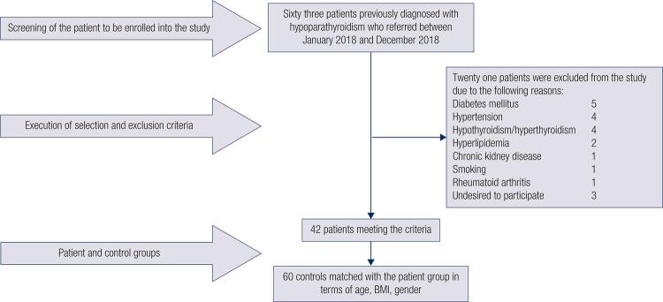
Objective The aim of the present study was to evaluate whether arterial stiffness is affected in the patients with hypoparathyroidism through pulse wave analysis (PWA). Subjects and methods Sixty-three patients diagnosed with hypoparathyroidism and sixty volunteers were evaluated for the study. When 21 patients were excluded in the hypoparathyroidism group due to exclusion criteria, the research continued with 42 patients and 60 volunteers who are similar to the patients in terms of age, gender and body mass index (BMI). Fasting plasma glucose after 10 hours of fasting, creatinine, thyroid stimulating hormone (TSH), free thyroxine (fT4), albumin, calcium, phosphorus, magnesium, 25-OH vitamin D, parathormone (PTH) and urine calcium results in 24-hour urine for the patients in the hypoparathyroidism group were recorded. Evaluation of arterial stiffness was performed by Mobil-O-Graph 24h PWA device. Results Systolic blood pressure (SBP) (p = 0.01), diastolic blood pressure (DBP) (p = 0.005), mean blood pressure (p = 0.009), central SBP (p = 0.004), central DBP (p = 0.01) and pulse wave velocity (PWV) (p = 0.02) were found higher in the hypoparathyroidism group. A positive correlation was detected between phosphorus level and SBP [(p = 0.03. r = 0.327)], central SBP [(p = 0.04, r = 0.324)] and PWV [(p = 0.003, r = 0.449)]. We detected that age and serum phosphorus levels were independent predictor variables for PWV (B = 0.014, p < 0.001 and B = 0.035, p < 0.001, respectively). Conclusion We detected that hypoparathyroidism causes an increase in blood pressure and arterial stiffness. The most significant determinant factors were detected as advanced age and hyperphosphatemia. The patients diagnosed with hypoparathyroidism should be closely monitored and treatment planning should include to prevent the patients from hyperphosphatemia.
目的 本研究旨在通过脉搏波分析(PWA)评估甲状旁腺功能减退患者的动脉僵硬度是否受到影响。
受试者与方法 对63例诊断为甲状旁腺功能减退的患者和60名志愿者进行了研究评估。由于排除标准,甲状旁腺功能减退组中有21例患者被排除,研究继续对42例患者和60名在年龄、性别和体重指数(BMI)方面与患者相似的志愿者进行。记录甲状旁腺功能减退组患者禁食10小时后的空腹血糖、肌酐、促甲状腺激素(TSH)、游离甲状腺素(fT4)、白蛋白、钙、磷、镁、25-羟基维生素D、甲状旁腺激素(PTH)以及24小时尿钙结果。使用Mobil-O-Graph 24小时PWA设备进行动脉僵硬度评估。
结果 甲状旁腺功能减退组的收缩压(SBP)(p = 0.01)、舒张压(DBP)(p = 0.005)、平均血压(p = 0.009)、中心SBP(p = 0.004)、中心DBP(p = 0.01)和脉搏波速度(PWV)(p = 0.02)均较高。发现磷水平与SBP[(p = 0.03,r = 0.327)]、中心SBP[(p = 0.04,r = 0.324)]和PWV[(p = 0.003,r = 0.449)]之间存在正相关。我们检测到年龄和血清磷水平是PWV的独立预测变量(分别为B = 0.014,p < 0.001和B = 0.035,p < 0.001)。
结论 我们检测到甲状旁腺功能减退会导致血压和动脉僵硬度增加。检测到最显著的决定因素是高龄和高磷血症。对诊断为甲状旁腺功能减退的患者应密切监测,治疗计划应包括预防患者出现高磷血症。