• 文献检索
  • 文档翻译
  • 深度研究
  • 学术资讯
  • Suppr Zotero 插件Zotero 插件
  • 邀请有礼
  • 套餐&价格
  • 历史记录
应用&插件
Suppr Zotero 插件Zotero 插件浏览器插件Mac 客户端Windows 客户端微信小程序
定价
高级版会员购买积分包购买API积分包
服务
文献检索文档翻译深度研究API 文档MCP 服务
关于我们
关于 Suppr公司介绍联系我们用户协议隐私条款
关注我们

Suppr 超能文献

核心技术专利:CN118964589B侵权必究
粤ICP备2023148730 号-1Suppr @ 2026

文献检索

告别复杂PubMed语法,用中文像聊天一样搜索,搜遍4000万医学文献。AI智能推荐,让科研检索更轻松。

立即免费搜索

文件翻译

保留排版,准确专业,支持PDF/Word/PPT等文件格式,支持 12+语言互译。

免费翻译文档

深度研究

AI帮你快速写综述,25分钟生成高质量综述,智能提取关键信息,辅助科研写作。

立即免费体验

基于影像学的骨密度和骨质量测量的最新进展。

Update on Imaging-Based Measurement of Bone Mineral Density and Quality.

机构信息

Department of Radiology, University of California, 400 Parnassus Ave, A-367, San Francisco, CA, 94143, USA.

出版信息

Curr Rheumatol Rep. 2020 Apr 9;22(5):13. doi: 10.1007/s11926-020-00892-w.

DOI:10.1007/s11926-020-00892-w
PMID:32270332
原文链接:https://pmc.ncbi.nlm.nih.gov/articles/PMC7875476/
Abstract

PURPOSE OF REVIEW

Patients with inflammatory arthropathies have a high rate of fragility fractures. Diagnostic assessment and monitoring of bone density and quality are therefore critically important. Here, we review standard and advanced techniques to measure bone density and quality, specifically focusing on patients with inflammatory arthropathies.

RECENT FINDINGS

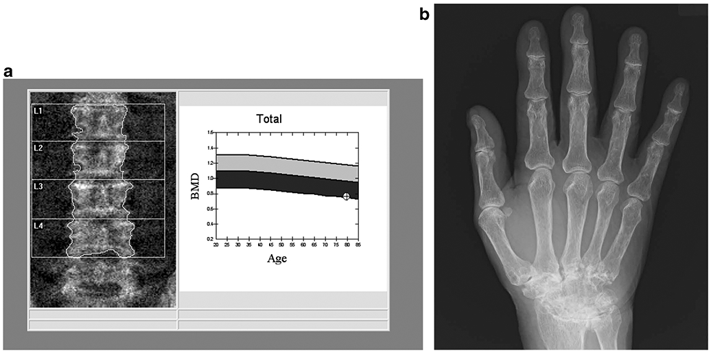
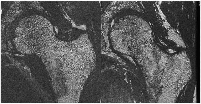
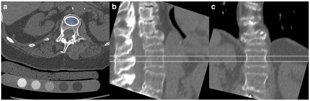
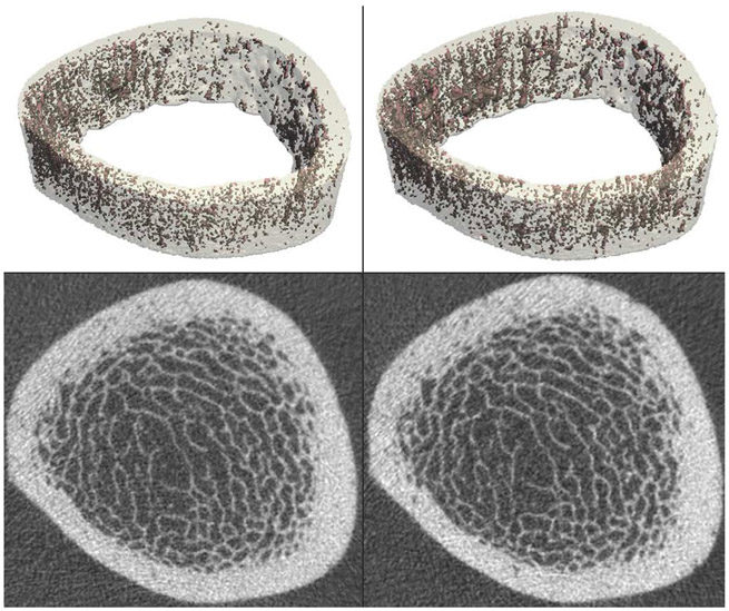
Current standard procedures are dual-energy X-ray absorptiometry (DXA) and quantitative computed tomography (QCT). DXA-based newer methods include trabecular bone score (TBS) and vertebral fracture assessment (VFA). More advanced imaging methods to measure bone quality include high-resolution peripheral quantitative computed tomography (HR-pQCT) as well as multi-detector CT (MD-CT) and magnetic resonance imaging (MRI). Quantitative ultrasound has shown promise but is not standard to assess bone fragility. While there are limitations, DXA remains the standard technique to measure density in patients with rheumatological disorders. Newer modalities to measure bone quality may allow better characterization of bone fragility but currently are not standard of care procedures.

摘要

目的综述

炎症性关节炎患者脆性骨折的发生率很高。因此,对骨密度和骨质量进行诊断评估和监测至关重要。在这里,我们综述了评估骨密度和骨质量的标准和先进技术,特别是针对炎症性关节炎患者。

最新发现

目前的标准方法是双能 X 射线吸收法(DXA)和定量计算机断层扫描(QCT)。基于 DXA 的新方法包括骨小梁评分(TBS)和椎体骨折评估(VFA)。评估骨质量的更先进的成像方法包括高分辨率外周定量计算机断层扫描(HR-pQCT)以及多探测器 CT(MD-CT)和磁共振成像(MRI)。定量超声显示出一定的前景,但不是评估骨脆性的标准方法。虽然存在局限性,但 DXA 仍然是评估风湿性疾病患者骨密度的标准技术。测量骨质量的新方法可能可以更好地描述骨脆性,但目前还不是常规的护理程序。

https://cdn.ncbi.nlm.nih.gov/pmc/blobs/63df/7875476/0e449b51ce2c/nihms-1664770-f0004.jpg
https://cdn.ncbi.nlm.nih.gov/pmc/blobs/63df/7875476/66bf0b175214/nihms-1664770-f0001.jpg
https://cdn.ncbi.nlm.nih.gov/pmc/blobs/63df/7875476/72da78ecaf38/nihms-1664770-f0002.jpg
https://cdn.ncbi.nlm.nih.gov/pmc/blobs/63df/7875476/f822742c59ed/nihms-1664770-f0003.jpg
https://cdn.ncbi.nlm.nih.gov/pmc/blobs/63df/7875476/0e449b51ce2c/nihms-1664770-f0004.jpg

相似文献

1
Update on Imaging-Based Measurement of Bone Mineral Density and Quality.基于影像学的骨密度和骨质量测量的最新进展。
Curr Rheumatol Rep. 2020 Apr 9;22(5):13. doi: 10.1007/s11926-020-00892-w.
2
Imaging in osteogenesis imperfecta: Where we are and where we are going.成骨不全症的影像学表现:现状与未来。
Eur J Med Genet. 2024 Apr;68:104926. doi: 10.1016/j.ejmg.2024.104926. Epub 2024 Feb 16.
3
Changes in cortical microarchitecture are independent of areal bone mineral density in patients with fragility fractures.脆性骨折患者皮质微结构的变化与局部骨矿物质密度无关。
Injury. 2017 Nov;48(11):2461-2465. doi: 10.1016/j.injury.2017.08.043. Epub 2017 Aug 25.
4
Vertebral fracture assessment by dual-energy X-ray absorptiometry along with bone mineral density in the evaluation of postmenopausal osteoporosis.双能 X 射线吸收法评估椎体骨折联合骨密度在绝经后骨质疏松症评估中的应用。
Arch Osteoporos. 2020 Feb 24;15(1):25. doi: 10.1007/s11657-020-0688-9.
5
Peripheral Quantitative Computed Tomography (pQCT) Measures Contribute to the Understanding of Bone Fragility in Older Patients With Low-trauma Fracture.外周定量计算机断层扫描(pQCT)测量有助于理解低创伤骨折老年患者的骨脆性。
J Clin Densitom. 2018 Jan-Mar;21(1):140-147. doi: 10.1016/j.jocd.2017.02.003. Epub 2017 Mar 9.
6
"Which skeletal imaging modality is best for assessing bone health in children and young adults compared to DXA? A systematic review and meta-analysis".“与 DXA 相比,哪种骨骼成像方式最适合评估儿童和青少年的骨骼健康?系统评价和荟萃分析”。
Bone. 2021 Sep;150:116013. doi: 10.1016/j.bone.2021.116013. Epub 2021 May 23.
7
Quantitative computed tomography discriminates between postmenopausal women with low spine bone mineral density with vertebral fractures and those with low spine bone mineral density only: the SHATTER study.定量计算机断层扫描可区分伴有椎体骨折的绝经后低骨密度女性和仅有低骨密度的绝经后女性:SHATTER 研究。
Osteoporos Int. 2020 Apr;31(4):667-675. doi: 10.1007/s00198-020-05317-z. Epub 2020 Jan 28.
8
Association between bone indices assessed by DXA, HR-pQCT and QCT scans in post-menopausal women.双能X线吸收法(DXA)、高分辨率外周定量CT(HR-pQCT)和定量CT(QCT)扫描评估的绝经后女性骨指标之间的关联。
J Bone Miner Metab. 2016 Nov;34(6):638-645. doi: 10.1007/s00774-015-0708-9. Epub 2015 Aug 21.
9
Vertebral bone attenuation on low-dose chest CT: quantitative volumetric analysis for bone fragility assessment.低剂量胸部CT上的椎体骨质衰减:用于评估骨脆性的定量容积分析
Osteoporos Int. 2017 Jan;28(1):329-338. doi: 10.1007/s00198-016-3724-2. Epub 2016 Aug 1.
10
Associations between radius low-frequency axial ultrasound velocity and bone fragility in elderly men and women.老年人桡骨低频轴向超声速度与骨脆性的相关性研究。
Osteoporos Int. 2019 Feb;30(2):411-421. doi: 10.1007/s00198-018-4725-0. Epub 2018 Oct 10.

引用本文的文献

1
Comparison of thoracic vertebrae and L1 CT attenuation in predicting osteoporosis using opportunistic chest CT.利用机会性胸部CT比较胸椎和L1的CT衰减值在预测骨质疏松症中的作用
Ther Adv Musculoskelet Dis. 2025 Sep 11;17:1759720X251374134. doi: 10.1177/1759720X251374134. eCollection 2025.
2
Opportunistic osteoporosis assessment from routine CT-effect of intravenous contrast agents on absolute values, T-scores, and derived classifications in single- and dual-energy CT.基于常规CT的机会性骨质疏松评估——静脉造影剂对单能和双能CT中绝对值、T值及衍生分类的影响
Eur Radiol. 2025 Sep 11. doi: 10.1007/s00330-025-11988-1.
3

本文引用的文献

1
Detection of Atypical Femur Fractures.检测非典型股骨骨折。
J Clin Densitom. 2019 Oct-Dec;22(4):506-516. doi: 10.1016/j.jocd.2019.07.003. Epub 2019 Jul 10.
2
The association between prevalent vertebral fractures and bone quality of the distal radius and distal tibia as measured with HR-pQCT in postmenopausal women with a recent non-vertebral fracture at the Fracture Liaison Service.在骨折联络服务处,对近期发生过非椎体骨折的绝经后女性,用 HR-pQCT 测量桡骨远端和胫骨远端的骨质量,研究其与普遍存在的椎体骨折之间的相关性。
Osteoporos Int. 2019 Sep;30(9):1789-1797. doi: 10.1007/s00198-019-05081-9. Epub 2019 Jul 17.
3
Multi-detector CT for suspected hip fragility fractures: A diagnostic test accuracy systematic review and meta-analysis.
Evaluation of bone mineral density in patients with cervical ossification of the posterior longitudinal ligament utilizing vertebral bone quality and Hounsfield units.
利用椎体骨质量和亨氏单位评估后纵韧带骨化症患者的骨密度
Eur Spine J. 2025 Aug 11. doi: 10.1007/s00586-025-09212-1.
4
Research on the predictive efficacy of MRI-based endplate bone quality and vertebral body bone quality scores for adjacent segment degeneration after lumbar spinal fusion.基于MRI的终板骨质量和椎体骨质量评分对腰椎融合术后相邻节段退变的预测效能研究
Eur Spine J. 2025 Aug 9. doi: 10.1007/s00586-025-09239-4.
5
Opportunistic computed tomography (CT) assessment of osteoporosis in patients undergoing transcatheter aortic valve replacement (TAVR).经导管主动脉瓣置换术(TAVR)患者骨质疏松的机会性计算机断层扫描(CT)评估
Arch Osteoporos. 2025 Jul 17;20(1):100. doi: 10.1007/s11657-025-01579-4.
6
Finite element fracture load analysis and dark-field X-ray imaging of osteoporotic and healthy vertebrae in human lumbar spine specimens.人腰椎标本中骨质疏松性和健康椎体的有限元骨折载荷分析及暗场X射线成像
BMC Musculoskelet Disord. 2025 Jun 3;26(1):545. doi: 10.1186/s12891-025-08709-6.
7
Comparative evaluation of accuracy, precision, and radiation dose between mindways and low-dose iCare QCT for lumbar spine BMD using the European spine phantom.使用欧洲脊柱模型对Mindways和低剂量iCare QCT在腰椎骨密度测量方面的准确性、精密度和辐射剂量进行比较评估。
Front Med Technol. 2025 May 12;7:1575553. doi: 10.3389/fmedt.2025.1575553. eCollection 2025.
8
Prediction of fusion cage subsidence after transforaminal lumbar interbody fusion based on MRI endplate bone quality.基于MRI终板骨质质量预测经椎间孔腰椎椎间融合术后融合器下沉
Quant Imaging Med Surg. 2025 May 1;15(5):4122-4134. doi: 10.21037/qims-20204-2533. Epub 2025 Apr 24.
9
Simulated low-dose multi-detector computed tomography: spatial effects on surrogate parameters of bone strength at the proximal femur.模拟低剂量多探测器计算机断层扫描:对股骨近端骨强度替代参数的空间影响。
Osteoporos Int. 2025 May;36(5):917-928. doi: 10.1007/s00198-025-07467-4. Epub 2025 Apr 5.
10
Generate Quantitative Computed Tomography-Equivalent Computed Tomography Image Interpretation Reports in Patients with Spinal Deformities.生成脊柱畸形患者的定量计算机断层扫描等效计算机断层扫描图像解读报告。
J Bone Metab. 2025 Feb;32(1):31-37. doi: 10.11005/jbm.24.801. Epub 2025 Feb 28.
多层螺旋CT用于疑似髋部脆性骨折:一项诊断试验准确性的系统评价和荟萃分析。
Emerg Radiol. 2019 Oct;26(5):549-556. doi: 10.1007/s10140-019-01696-x. Epub 2019 Jun 17.
4
Bone Mineral Density and Microstructure of the Elbow in Baseball Pitchers: An Analysis by Second-Generation HR-pQCT.棒球投手肘部的骨密度和微观结构:第二代 HR-pQCT 分析。
J Clin Densitom. 2020 Apr-Jun;23(2):322-328. doi: 10.1016/j.jocd.2019.03.001. Epub 2019 Mar 16.
5
Pediatric bone evaluation with HR-pQCT: A comparison between standard and height-adjusted positioning protocols in a cohort of teenagers with chronic kidney disease.采用高分辨率外周定量CT进行儿童骨骼评估:慢性肾病青少年队列中标准定位方案与身高校正定位方案的比较
Arch Pediatr. 2019 Apr;26(3):151-157. doi: 10.1016/j.arcped.2019.02.003. Epub 2019 Mar 1.
6
Longitudinal changes in subchondral bone structure as assessed with MRI are associated with functional outcome after high tibial osteotomy.通过MRI评估的胫骨近端截骨术后软骨下骨结构的纵向变化与功能结果相关。
J ISAKOS. 2018 Jul 20;3(4):205-212. doi: 10.1136/jisakos-2017-000194. Epub 2018 Jun 28.
7
3T chemical shift-encoded MRI: Detection of altered proximal femur marrow adipose tissue composition in glucocorticoid users and validation with magnetic resonance spectroscopy.3T 化学位移编码 MRI:检测糖皮质激素使用者股骨近端骨髓脂肪组织成分的改变,并通过磁共振波谱进行验证。
J Magn Reson Imaging. 2019 Aug;50(2):490-496. doi: 10.1002/jmri.26586. Epub 2018 Dec 12.
8
Cortical and trabecular bone microarchitecture as an independent predictor of incident fracture risk in older women and men in the Bone Microarchitecture International Consortium (BoMIC): a prospective study.皮质骨和小梁骨微结构作为骨微结构国际联合会(BoMIC)中老年男女新发骨折风险的独立预测因子:一项前瞻性研究。
Lancet Diabetes Endocrinol. 2019 Jan;7(1):34-43. doi: 10.1016/S2213-8587(18)30308-5. Epub 2018 Nov 28.
9
Prediction of incident vertebral fracture using CT-based finite element analysis.基于 CT 的有限元分析预测新发椎体骨折。
Osteoporos Int. 2019 Feb;30(2):323-331. doi: 10.1007/s00198-018-4716-1. Epub 2018 Oct 10.
10
Current imaging techniques in osteoporosis.骨质疏松症的当前影像学技术。
Clin Exp Rheumatol. 2018 Sep-Oct;36 Suppl 114(5):115-126. Epub 2018 Oct 1.