Department of Dermatology, Peking Union Medical College Hospital, Chinese Academy of Medical Sciences and Peking Union Medical College, Beijing, China.
National Clinical Research Center for Dermatologic and Immunologic Diseases, Beijing, China.
J Immunol Res. 2020 Mar 21;2020:5980190. doi: 10.1155/2020/5980190. eCollection 2020.
Psoriasis is an immune-mediated chronic inflammatory skin disorder in which the dysregulation of immune cells plays an important role in its development. Tumor necrosis factor- (TNF-) antagonists affect the immune repertoire, while TNF--induced protein 3 (TNFAIP3) has a protective role against the deleterious effects of inflammation and participates in immune regulation.
We investigated the immune regulation of in the pathogenesis of psoriasis and determined whether it is involved in the antipsoriatic effect of TNF- antagonists.
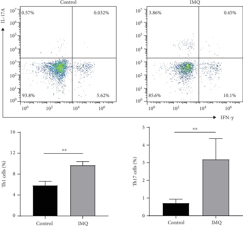
mRNA levels were evaluated in blood from patients with moderate-to-severe psoriasis. The effects of TNF- antagonists were examined in a mouse imiquimod- (IMQ-) induced psoriasis-like dermatitis model. In the mouse model, mRNA expression was determined using RT-PCR. Serum levels of IL-17A, IL-23, IFN-, TNF-, phosphorylated ERK1/2, p38, and JNK were measured using ELISA. The proportion of Th1 and Th17 cells in mouse spleens was analyzed using flow cytometry.
mRNA expression levels of in the blood were significantly lower in patients with moderate and severe psoriasis (mean ± SD = 0.44 ± 0.25) compared with normal subjects (mean ± SD = 1.00 ± 0.82) ( < 0.01). In the mouse model, IMQ downregulated expression levels, which were increased after TNF- antagonist treatment ( < 0.05). Serum levels of Th17 cytokines (IL-17A and IL-23) and Th1 cytokines (IFN- and TNF-) were significantly higher in the IMQ and IMQ/rat IgG1 groups compared with the control group, and the application of TNF- antagonists significantly decreased the levels of inflammatory cytokines ( < 0.01). Notably, phosphorylated p38 levels were increased in the IMQ and IMQ/rat IgG1 groups compared with the control group but were downregulated by treatment with TNF- antagonists ( < 0.05). Th1 and Th17 cells were significantly increased in the IMQ group compared with the control group ( < 0.01).
downregulation associated with Th1 and Th17 cell differentiation and p38 activation might contribute in part to the mechanism of immune dysfunction in psoriasis. TNF- antagonists might partly exert their effects on psoriasis via this pathway.
银屑病是一种免疫介导的慢性炎症性皮肤疾病,其中免疫细胞的失调在其发病机制中起着重要作用。肿瘤坏死因子-(TNF-)拮抗剂影响免疫谱,而 TNF--诱导蛋白 3(TNFAIP3)具有对抗炎症的有害影响的保护作用,并参与免疫调节。
我们研究了 在银屑病发病机制中的免疫调节作用,并确定其是否参与 TNF-拮抗剂的抗银屑病作用。
评估了中重度银屑病患者的血液中 mRNA 水平。在咪喹莫特(IMQ)诱导的银屑病样皮炎小鼠模型中研究了 TNF-拮抗剂的作用。在小鼠模型中,通过 RT-PCR 确定 mRNA 表达。使用 ELISA 测量血清中白细胞介素 17A(IL-17A)、白细胞介素 23(IL-23)、干扰素-(IFN-)、肿瘤坏死因子-(TNF-)、磷酸化 ERK1/2、p38 和 JNK 的水平。使用流式细胞术分析小鼠脾脏中 Th1 和 Th17 细胞的比例。
中重度银屑病患者(均值 ± SD=0.44±0.25)的血液中 mRNA 表达水平明显低于正常对照者(均值 ± SD=1.00±0.82)( < 0.01)。在小鼠模型中,IMQ 下调 表达水平,而 TNF-拮抗剂治疗后其表达水平增加( < 0.05)。与对照组相比,IMQ 和 IMQ/大鼠 IgG1 组的 Th17 细胞因子(IL-17A 和 IL-23)和 Th1 细胞因子(IFN-和 TNF-)的血清水平明显升高,而 TNF-拮抗剂的应用显著降低了炎症细胞因子的水平( < 0.01)。值得注意的是,与对照组相比,IMQ 和 IMQ/大鼠 IgG1 组的磷酸化 p38 水平升高,但经 TNF-拮抗剂治疗后下调( < 0.05)。与对照组相比,IMQ 组 Th1 和 Th17 细胞明显增加( < 0.01)。
与 Th1 和 Th17 细胞分化和 p38 激活相关的下调可能部分导致银屑病免疫功能障碍的机制。TNF-拮抗剂可能通过该途径部分发挥其对银屑病的作用。