Laboratory of Medical Genetics, Faculty of Biology and Environmental Protection, University of Lodz, Lodz, Poland.
Department of Medical Biochemistry, Medical University of Lodz, Lodz, Poland.
J Cell Mol Med. 2020 May;24(10):5675-5694. doi: 10.1111/jcmm.15231. Epub 2020 Apr 13.
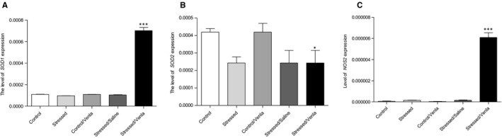
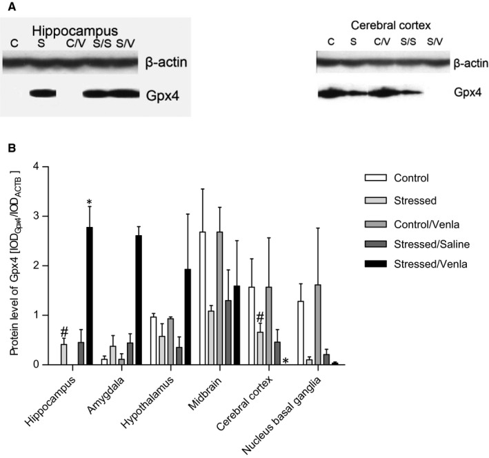
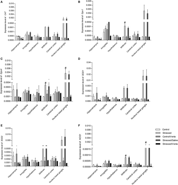
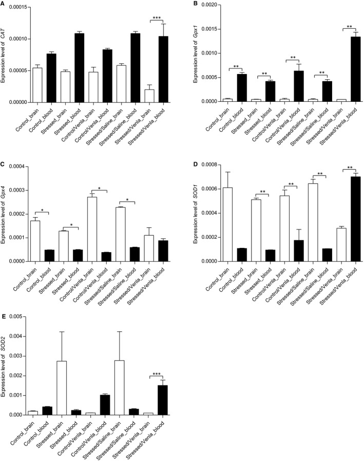
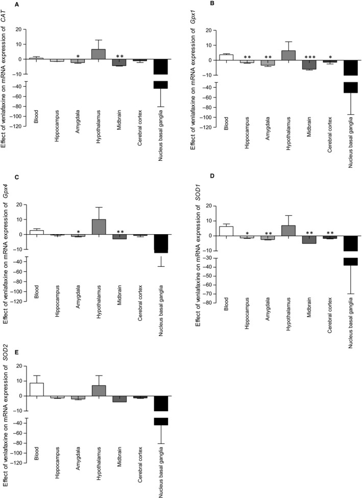
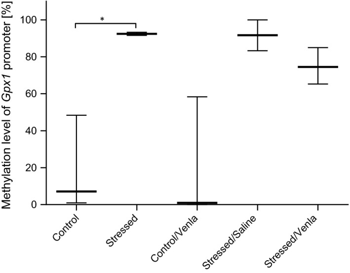
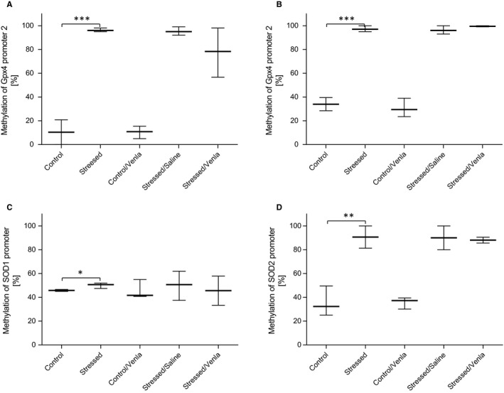
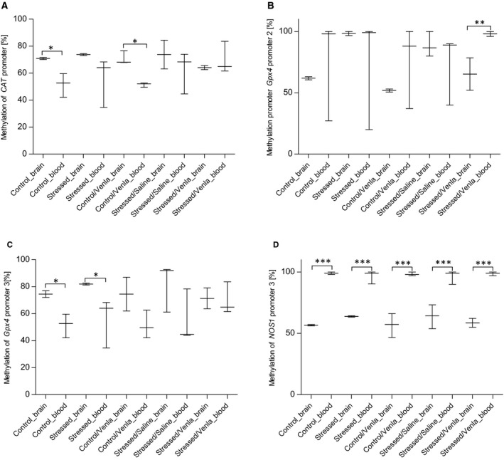
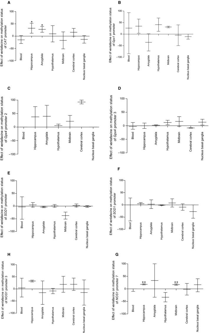
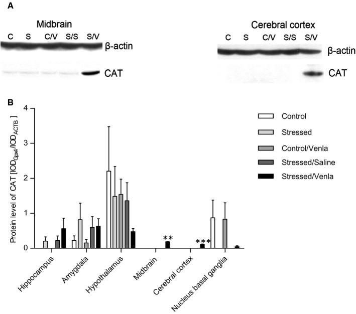
Recent human and animal studies indicate that oxidative and nitrosative stress may play a role in the aetiology and pathogenesis of depression. This study investigates the effect of chronic administration of the serotonin-norepinephrine reuptake inhibitor, venlafaxine, on the expression and methylation status of SOD1, SOD2, GPx1, GPx4, CAT, NOS1 and NOS2 in the brain and blood of rats exposed to a chronic mild stress (CMS) model of depression. Separate groups of animals were exposed to CMS for 2 or 7 weeks; the second group received saline or venlafaxine (10 mg/kg/d, IP) for 5 weeks. After completion of both stress conditions and drug administration, the mRNA and protein expression of selected genes and the methylation status of their promoters were measured in peripheral mononuclear blood cells (PBMCs) and in brain structures (hippocampus, amygdala, hypothalamus, midbrain, cortex, basal ganglia) with the use of TaqMan Gene Expression Assay, Western blot and methylation-sensitive high-resolution melting techniques. CMS caused a decrease in sucrose consumption, and this effect was normalized by fluoxetine. In PBMCs, SOD1, SOD2 and NOS2 mRNA expression changed only after venlafaxine administration. In brain, CAT, Gpx1, Gpx4 and NOS1 gene expression changed following CMS or venlafaxine exposure, most prominently in the hippocampus, midbrain and basal ganglia. CMS increased the methylation of the Gpx1 promoter in PBMCs, the second Gpx4 promoter in midbrain and basal ganglia, and SOD1 and SOD2 in hippocampus. The CMS animals treated with venlafaxine displayed a significantly higher CAT level in midbrain and cerebral cortex. CMS caused an elevation of Gpx4 in the hippocampus, which was lowered in cerebral cortex by venlafaxine. The results indicate that CMS and venlafaxine administration affect the methylation of promoters of genes involved in oxidative and nitrosative stress. They also indicate that peripheral and central tissue differ in their response to stress or antidepressant treatments. It is possible that that apart from DNA methylation, a crucial role of expression level of genes may be played by other forms of epigenetic regulation, such as histone modification or microRNA interference. These findings provide strong evidence for thesis that analysis of the level of mRNA and protein expression as well as the status of promoter methylation can help in understanding the pathomechanisms of mental diseases, including depression, and the mechanisms of action of drugs effective in their therapy.
最近的人体和动物研究表明,氧化应激和硝化应激可能在抑郁症的病因和发病机制中起作用。本研究探讨了慢性给予 5-羟色胺去甲肾上腺素再摄取抑制剂文拉法辛对慢性轻度应激(CMS)抑郁模型大鼠大脑和血液中 SOD1、SOD2、GPx1、GPx4、CAT、NOS1 和 NOS2 的表达和甲基化状态的影响。分别将动物暴露于 CMS 2 或 7 周;第二组接受生理盐水或文拉法辛(10mg/kg/d,IP)治疗 5 周。完成两种应激条件和药物治疗后,使用 TaqMan 基因表达分析、Western blot 和甲基化敏感高分辨率熔解技术测量外周单核血淋巴细胞(PBMCs)和大脑结构(海马体、杏仁核、下丘脑、中脑、皮质、基底神经节)中选定基因的 mRNA 和蛋白质表达及其启动子的甲基化状态。CMS 导致蔗糖消耗减少,氟西汀可使这种作用正常化。在 PBMCs 中,SOD1、SOD2 和 NOS2 的 mRNA 表达仅在文拉法辛给药后发生变化。在大脑中,CAT、Gpx1、Gpx4 和 NOS1 基因表达在 CMS 或文拉法辛暴露后发生变化,在海马体、中脑和基底神经节最为明显。CMS 增加了 PBMCs 中 Gpx1 启动子的甲基化、中脑和基底神经节中第二个 Gpx4 启动子的甲基化以及海马体中的 SOD1 和 SOD2 甲基化。用文拉法辛治疗的 CMS 动物在中脑和大脑皮质中的 CAT 水平显著升高。CMS 导致海马体中 Gpx4 升高,而文拉法辛降低了大脑皮质中的 Gpx4。结果表明,CMS 和文拉法辛给药会影响参与氧化和硝化应激的基因启动子的甲基化。它们还表明,外周组织和中枢组织对应激或抗抑郁治疗的反应不同。除 DNA 甲基化外,基因表达水平的其他形式的表观遗传调控(如组蛋白修饰或 microRNA 干扰)可能发挥关键作用。这些发现为以下观点提供了有力证据:分析 mRNA 和蛋白质表达水平以及启动子甲基化状态有助于理解包括抑郁症在内的精神疾病的发病机制以及有效治疗药物的作用机制。