Zhu Yuan-Zheng, Hu Xuan, Zhang Jing, Wang Zhao-Hui, Wu Shu, Yi Yang-Yan
From the Department of Plastic Surgery, The Second Affiliated Hospital of Nanchang University, Jiangxi, P.R. China.
Ann Plast Surg. 2020 May;84(5):602-607. doi: 10.1097/SAP.0000000000002357.
Preventing scar formation during wound healing has important clinical implications. Numerous studies have indicated that adipose-derived stem cell culture mediums, which are rich in cytokines and extracellular vesicles (EVs), regulate matrix remodeling and prevent scar formation after wound healing. Therefore, using a rabbit scar model, we tried to demonstrate which factor in adipose-derived stem cell culture mediums plays a major role in preventing scar formation (EVs or cytokines), as well as revealing the underlying mechanism.
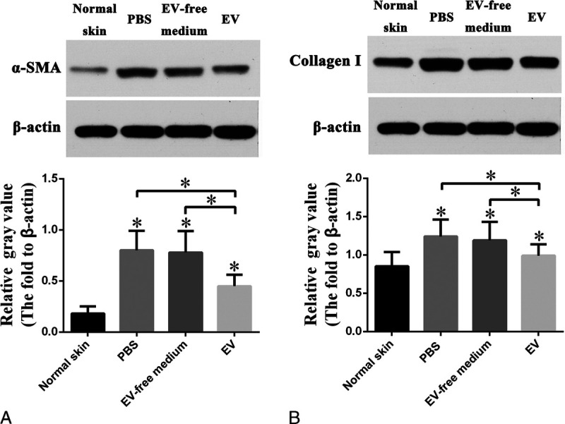
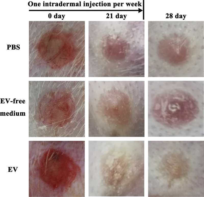
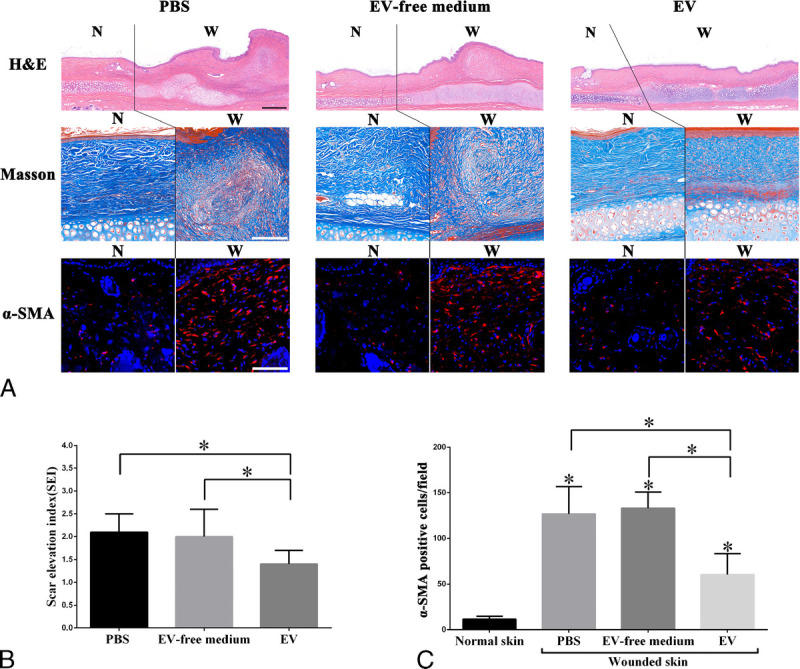
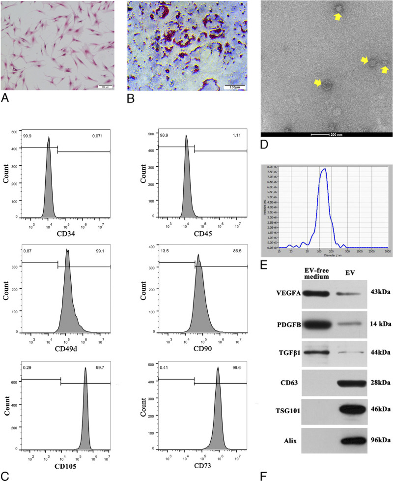
Human adipose-derived stem cells (hASCs) were isolated from the subcutaneous adipose tissue of a healthy female donor. The surface CD markers of third-passage hASCs were analyzed by flow cytometry. The adipogenic differentiation capacity of the hASCs was detected using Oil O staining. A cultured medium of third- to five-passage hASCs was collected for EV and EV-free medium isolations. Extracellular vesicles were characterized using transmission electron microscopy, NanoSight, and the Western blotting for surface markers CD63, TSG101, and Alix. The EV-free medium was characterized by Western blotting for vascular endothelial growth factor A (VEGFA), platelet derived growth factor B (PDGFB), and transforming growth factor β 1 (TGFβ1). Eight-millimeter-diameter wounds were created on the ventral side of both ears of 16 New Zealand rabbits. A total of 0.1 mL of the human adipose-derived stem cell-extracellular vesicle (hASC-EV) or EV-free medium was locally injected into wounds made on the right ears during wound healing. Meanwhile, equal amounts of phosphate buffer saline were injected into the left ears as a control. Biopsies of the wounded skin and surrounding tissue were excised on postoperative day 28 and subjected to hematoxylin and eosin (H&E), Masson, and α-SMA immunofluorescence staining. The protein expression of α-SMA and collagen I in both scar tissues and the normal skin were evaluated via Western blotting.
The hASCs expressed high levels of 49d, CD90, CD105, and CD73 but did not express CD34 or CD45. The hASCs differentiated into adipocytes under an adipogenic induction medium. Under transmission electron microscopy, the hASC-EVs were circular, bilayer membrane vesicles and approximately 95% of the particles were between 50 and 200 nm in size. The hASC-EVs expressed the same surface markers as EVs, including CD63, TSG101, and Alix and displayed little expression of VEGFA, PDGFB, and TGFβ1. The EV-free medium had a high expression of VEGFA, PDGFB, and TGFβ1 but displayed no expression of CD63, TSG101, and Alix. In vivo, the hASC-EV treatment prevented the formation of hypertrophic scars on postoperative day 28 and suppressed collagen deposition and myofibroblast aggregation. However, the EV-free medium did not prevent the formation of hypertrophic scars on the same time point and had little effect on collagen deposition and myofibroblast aggregation when compared with the control group.
Our study suggests that hASCs are associated with preventive scar formation therapy because of paracrine EVs rather than cytokines. A local injection of hASC-EVs during wound healing efficiently prevented hypertrophic scar formation, which may have a clinically beneficial antiscarring effect.
在伤口愈合过程中预防瘢痕形成具有重要的临床意义。大量研究表明,富含细胞因子和细胞外囊泡(EVs)的脂肪来源干细胞培养基可调节基质重塑并预防伤口愈合后的瘢痕形成。因此,我们利用兔瘢痕模型,试图证明脂肪来源干细胞培养基中的哪种因素在预防瘢痕形成中起主要作用(EVs还是细胞因子),并揭示其潜在机制。
从一名健康女性供体的皮下脂肪组织中分离出人脂肪来源干细胞(hASCs)。通过流式细胞术分析第三代hASCs的表面CD标志物。使用油红O染色检测hASCs的成脂分化能力。收集第三代至第五代hASCs的培养基用于分离EV和无EV培养基。使用透射电子显微镜、纳米可视技术以及针对表面标志物CD63、TSG101和Alix的蛋白质印迹法对细胞外囊泡进行表征。通过针对血管内皮生长因子A(VEGFA)、血小板衍生生长因子B(PDGFB)和转化生长因子β1(TGFβ1)的蛋白质印迹法对无EV培养基进行表征。在16只新西兰兔的双耳腹侧制造直径为8毫米的伤口。在伤口愈合期间,将总共0.1毫升的人脂肪来源干细胞细胞外囊泡(hASC-EV)或无EV培养基局部注射到右耳的伤口中。同时,将等量的磷酸盐缓冲盐水注射到左耳作为对照。在术后第28天切除受伤皮肤及周围组织的活检标本,并进行苏木精和伊红(H&E)染色、Masson染色以及α-SMA免疫荧光染色。通过蛋白质印迹法评估瘢痕组织和正常皮肤中α-SMA和胶原蛋白I的蛋白质表达。
hASCs高表达CD49d、CD90、CD105和CD73,但不表达CD34或CD45。在成脂诱导培养基下,hASCs分化为脂肪细胞。在透射电子显微镜下,hASC-EVs为圆形、双层膜囊泡,约95%的颗粒大小在50至200纳米之间。hASC-EVs表达与EVs相同的表面标志物,包括CD63、TSG101和Alix,并且VEGFA、PDGFB和TGFβ1的表达很少。无EV培养基中VEGFA、PDGFB和TGFβ1表达较高,但未检测到CD63、TSG101和Alix的表达。在体内,hASC-EV治疗可预防术后第28天肥厚性瘢痕的形成,并抑制胶原蛋白沉积和成肌纤维细胞聚集。然而,在同一时间点,无EV培养基并不能预防肥厚性瘢痕的形成,与对照组相比,对胶原蛋白沉积和成肌纤维细胞聚集几乎没有影响。
我们的研究表明,hASCs与预防性瘢痕形成治疗相关是由于其旁分泌的EVs而非细胞因子。在伤口愈合期间局部注射hASC-EVs可有效预防肥厚性瘢痕的形成,这可能具有临床有益的抗瘢痕作用。