• 文献检索
  • 文档翻译
  • 深度研究
  • 学术资讯
  • Suppr Zotero 插件Zotero 插件
  • 邀请有礼
  • 套餐&价格
  • 历史记录
应用&插件
Suppr Zotero 插件Zotero 插件浏览器插件Mac 客户端Windows 客户端微信小程序
定价
高级版会员购买积分包购买API积分包
服务
文献检索文档翻译深度研究API 文档MCP 服务
关于我们
关于 Suppr公司介绍联系我们用户协议隐私条款
关注我们

Suppr 超能文献

核心技术专利:CN118964589B侵权必究
粤ICP备2023148730 号-1Suppr @ 2026

文献检索

告别复杂PubMed语法,用中文像聊天一样搜索,搜遍4000万医学文献。AI智能推荐,让科研检索更轻松。

立即免费搜索

文件翻译

保留排版,准确专业,支持PDF/Word/PPT等文件格式,支持 12+语言互译。

免费翻译文档

深度研究

AI帮你快速写综述,25分钟生成高质量综述,智能提取关键信息,辅助科研写作。

立即免费体验

人朊病毒纤维的冷冻电镜结构,具有疏水性、抗蛋白酶的核心。

Cryo-EM structure of a human prion fibril with a hydrophobic, protease-resistant core.

机构信息

Department of Chemistry and Biochemistry; UCLA-DOE Institute for Genomics and Proteomics; STROBE, NSF Science and Technology Center, University of California, Los Angeles, Los Angeles, CA, USA.

Department of Biological Chemistry and Department of Chemistry and Biochemistry, UCLA-DOE Institute for Genomics and Proteomics, Howard Hughes Medical Institute, University of California Los Angeles, Los Angeles, CA, USA.

出版信息

Nat Struct Mol Biol. 2020 May;27(5):417-423. doi: 10.1038/s41594-020-0403-y. Epub 2020 Apr 13.

DOI:10.1038/s41594-020-0403-y
PMID:32284600
原文链接:https://pmc.ncbi.nlm.nih.gov/articles/PMC7338044/
Abstract

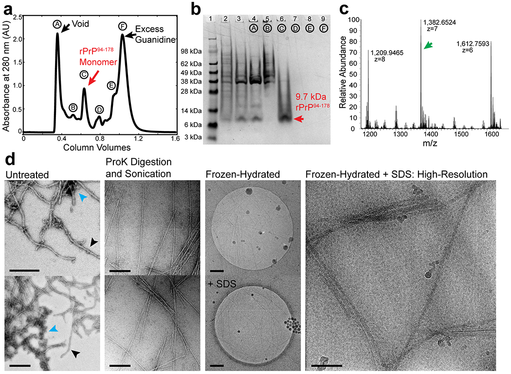
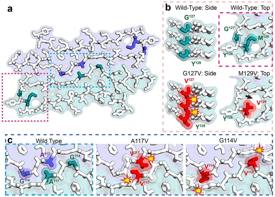
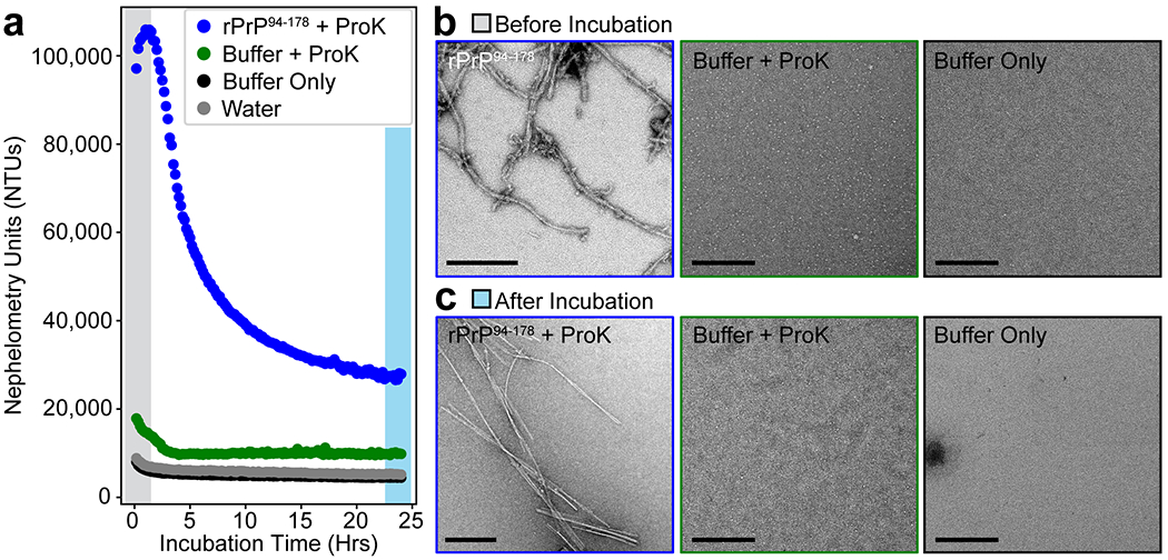
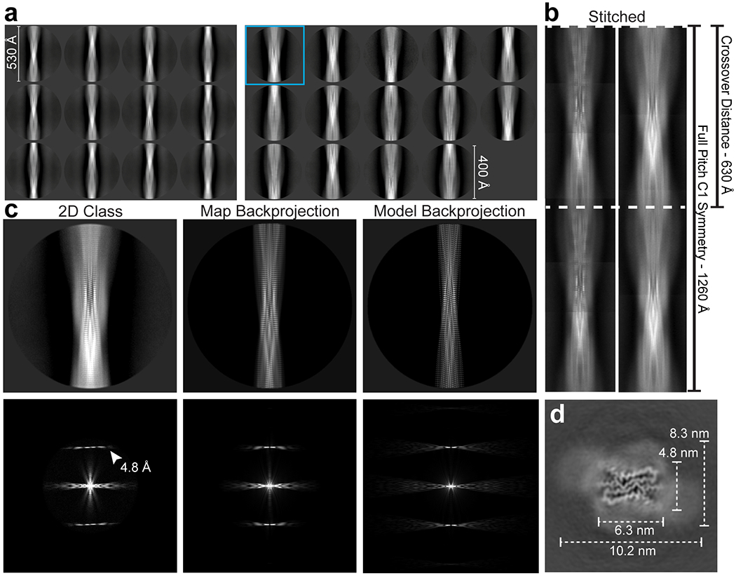
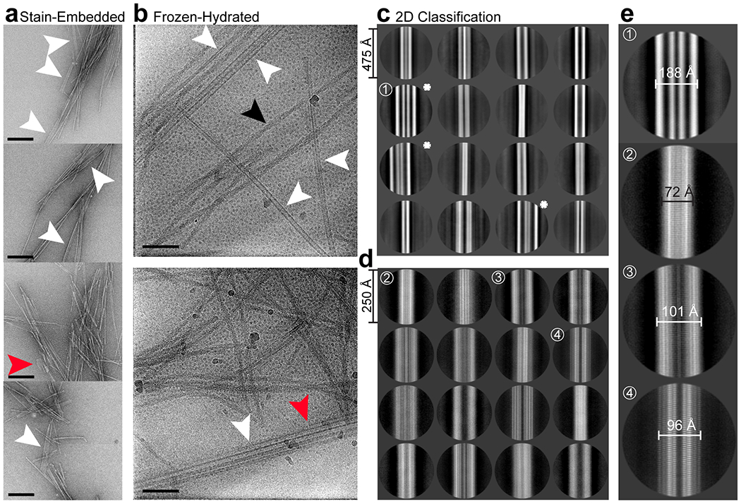
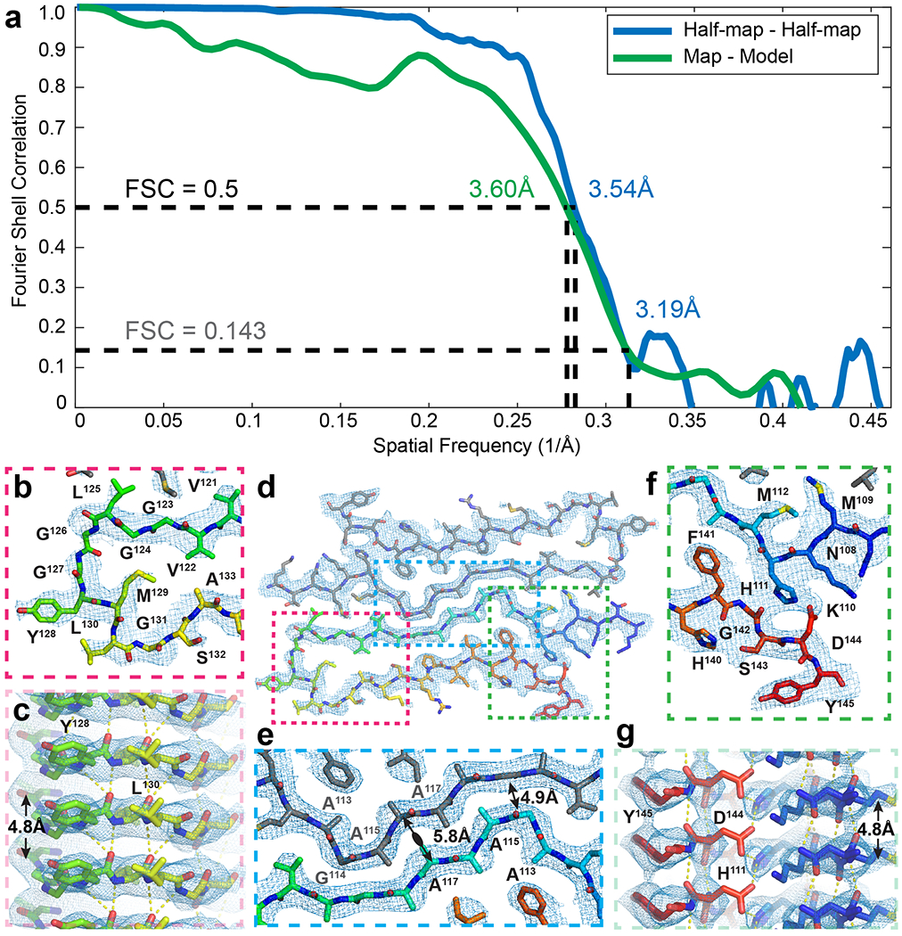
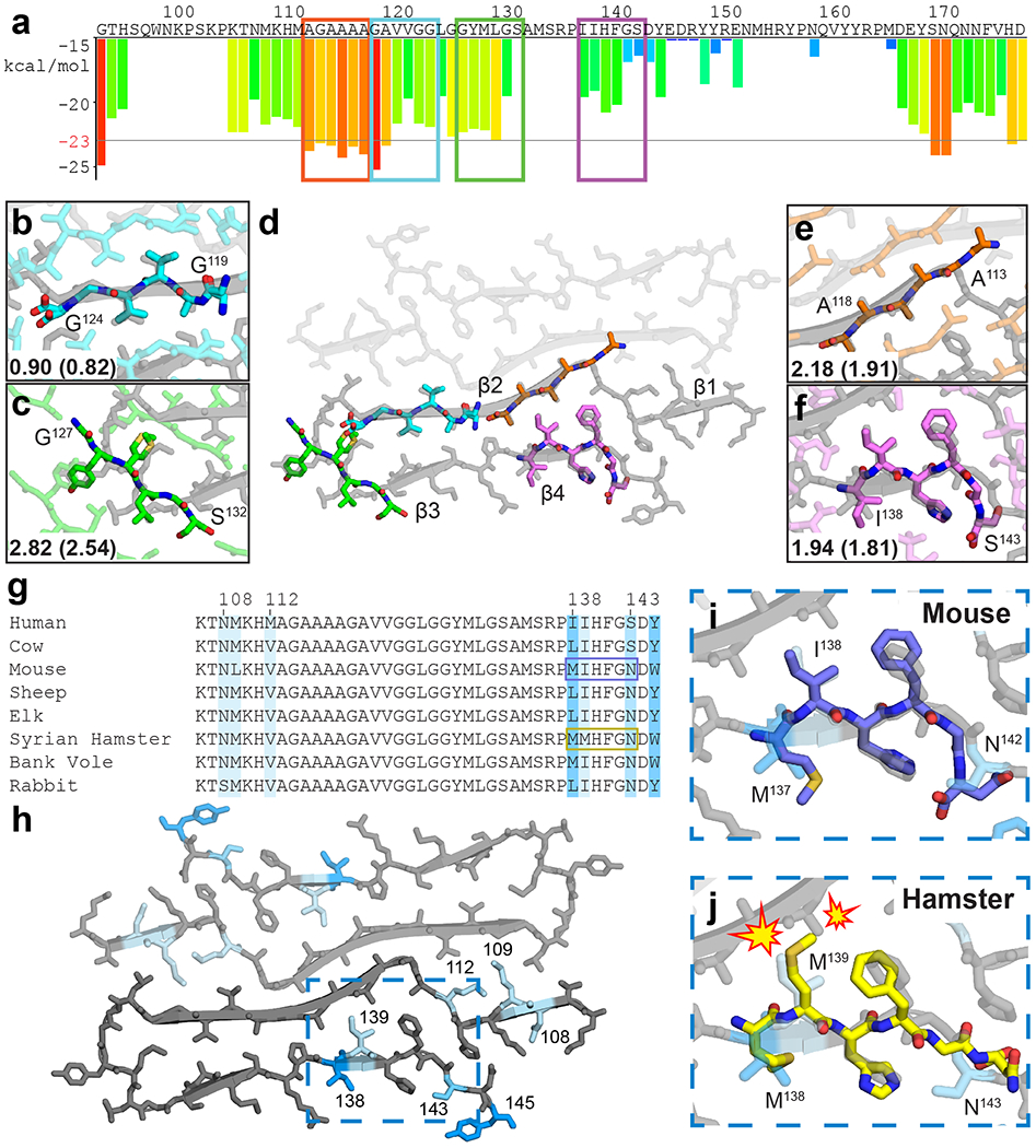
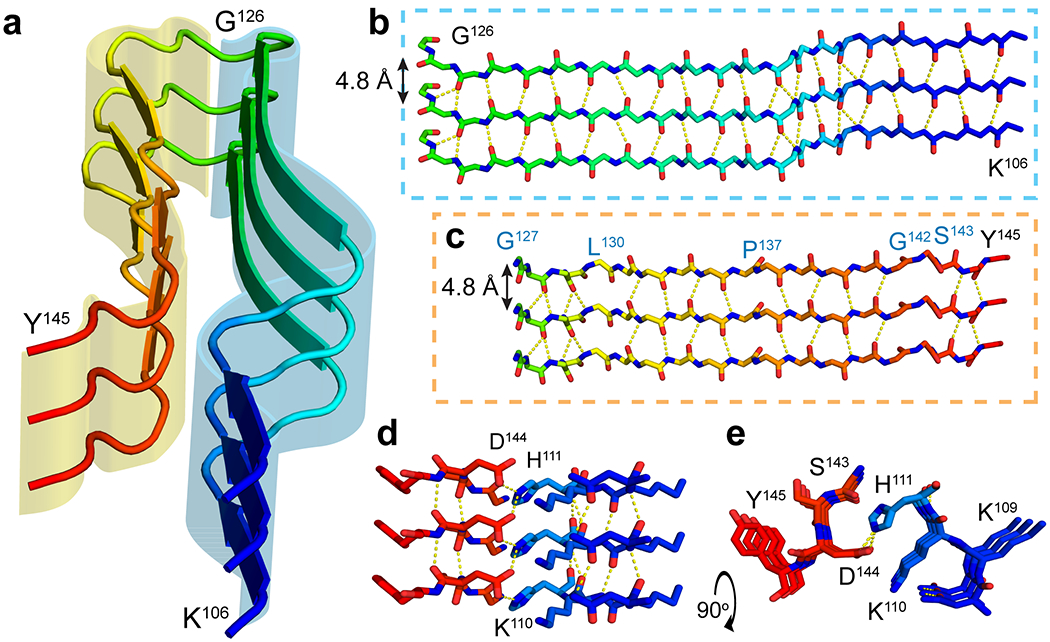
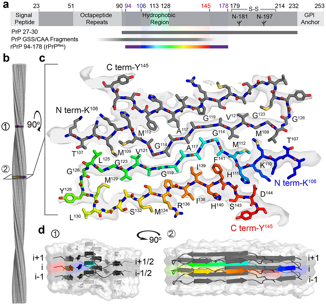
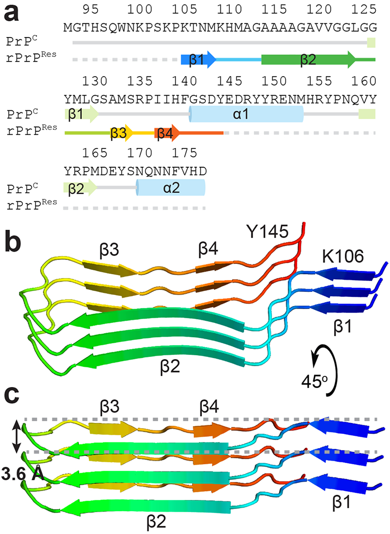
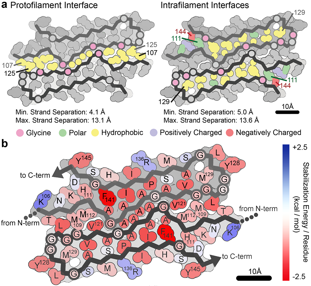
Self-templating assemblies of the human prion protein are clinically associated with transmissible spongiform encephalopathies. Here we present the cryo-EM structure of a denaturant- and protease-resistant fibril formed in vitro spontaneously by a 9.7-kDa unglycosylated fragment of the human prion protein. This human prion fibril contains two protofilaments intertwined with screw symmetry and linked by a tightly packed hydrophobic interface. Each protofilament consists of an extended beta arch formed by residues 106 to 145 of the prion protein, a hydrophobic and highly fibrillogenic disease-associated segment. Such structures of prion polymorphs serve as blueprints on which to evaluate the potential impact of sequence variants on prion disease.

摘要

朊病毒蛋白的自模板组装与可传播海绵状脑病在临床上有关。在这里,我们展示了一种在体外自发形成的变性剂和蛋白酶抗性纤维的冷冻电镜结构,它由人类朊病毒蛋白的 9.7kDa 非糖基化片段组成。这种人类朊病毒纤维包含两个原纤维,它们通过螺旋对称性相互交织,并通过紧密堆积的疏水性界面连接。每个原纤维由由朊病毒蛋白的 106 到 145 位残基形成的伸展β拱组成,这是一个疏水性和高度纤维原性的疾病相关片段。这些朊病毒多态体的结构可以作为蓝图,评估序列变异对朊病毒病的潜在影响。

https://cdn.ncbi.nlm.nih.gov/pmc/blobs/452e/7338044/ba68c6565e73/nihms-1569853-f0004.jpg
https://cdn.ncbi.nlm.nih.gov/pmc/blobs/452e/7338044/37d811f1161c/nihms-1569853-f0005.jpg
https://cdn.ncbi.nlm.nih.gov/pmc/blobs/452e/7338044/9db79892a75a/nihms-1569853-f0006.jpg
https://cdn.ncbi.nlm.nih.gov/pmc/blobs/452e/7338044/3d5b8ae9bcc6/nihms-1569853-f0007.jpg
https://cdn.ncbi.nlm.nih.gov/pmc/blobs/452e/7338044/c3d2c1eda42e/nihms-1569853-f0008.jpg
https://cdn.ncbi.nlm.nih.gov/pmc/blobs/452e/7338044/216a3b25d0ec/nihms-1569853-f0009.jpg
https://cdn.ncbi.nlm.nih.gov/pmc/blobs/452e/7338044/4639ab4fdd1f/nihms-1569853-f0010.jpg
https://cdn.ncbi.nlm.nih.gov/pmc/blobs/452e/7338044/00a94240a58f/nihms-1569853-f0011.jpg
https://cdn.ncbi.nlm.nih.gov/pmc/blobs/452e/7338044/4b59195b3c77/nihms-1569853-f0012.jpg
https://cdn.ncbi.nlm.nih.gov/pmc/blobs/452e/7338044/83bed26c4215/nihms-1569853-f0001.jpg
https://cdn.ncbi.nlm.nih.gov/pmc/blobs/452e/7338044/fe4d43fcb199/nihms-1569853-f0002.jpg
https://cdn.ncbi.nlm.nih.gov/pmc/blobs/452e/7338044/48e51f4b3099/nihms-1569853-f0003.jpg
https://cdn.ncbi.nlm.nih.gov/pmc/blobs/452e/7338044/ba68c6565e73/nihms-1569853-f0004.jpg

相似文献

1
Cryo-EM structure of a human prion fibril with a hydrophobic, protease-resistant core.人朊病毒纤维的冷冻电镜结构,具有疏水性、抗蛋白酶的核心。
Nat Struct Mol Biol. 2020 May;27(5):417-423. doi: 10.1038/s41594-020-0403-y. Epub 2020 Apr 13.
2
Self-Replication of Prion Protein Fragment 89-230 Amyloid Fibrils Accelerated by Prion Protein Fragment 107-143 Aggregates.朊病毒蛋白片段107 - 143聚集体加速朊病毒蛋白片段89 - 230淀粉样原纤维的自我复制
Int J Mol Sci. 2020 Oct 8;21(19):7410. doi: 10.3390/ijms21197410.
3
Pathogenic mutations within the hydrophobic domain of the prion protein lead to the formation of protease-sensitive prion species with increased lethality.朊病毒蛋白疏水结构域内的致病性突变会导致形成具有更高致死率的蛋白酶敏感型朊病毒。
J Virol. 2014 Mar;88(5):2690-703. doi: 10.1128/JVI.02720-13. Epub 2013 Dec 18.
4
Contribution of specific residues of the β-solenoid fold to HET-s prion function, amyloid structure and stability.β-螺旋折叠的特定残基对HET-s朊病毒功能、淀粉样结构和稳定性的贡献。
PLoS Pathog. 2014 Jun 12;10(6):e1004158. doi: 10.1371/journal.ppat.1004158. eCollection 2014 Jun.
5
Molecular structures of amyloid and prion fibrils: consensus versus controversy.淀粉样纤维和朊病毒纤维的分子结构:共识与争议。
Acc Chem Res. 2013 Jul 16;46(7):1487-96. doi: 10.1021/ar300282r. Epub 2013 Jan 7.
6
2.2 Å Cryo-EM Tetra-Protofilament Structure of the Hamster Prion 108-144 Fibril Reveals an Ordered Water Channel in the Center.2.2 Å Cryo-EM 四聚原纤维结构的仓鼠朊病毒 108-144 纤维揭示了中心的有序水通道。
J Am Chem Soc. 2022 Aug 3;144(30):13888-13894. doi: 10.1021/jacs.2c05479. Epub 2022 Jul 20.
7
2.7 Å cryo-EM structure of ex vivo RML prion fibrils.2.7 Å 冷冻电镜结构的 RML 朊病毒纤维。
Nat Commun. 2022 Jul 13;13(1):4004. doi: 10.1038/s41467-022-30457-7.
8
Cryo-EM structure of an amyloid fibril formed by full-length human prion protein.全长人类朊病毒蛋白形成的淀粉样纤维的冷冻电镜结构。
Nat Struct Mol Biol. 2020 Jun;27(6):598-602. doi: 10.1038/s41594-020-0441-5. Epub 2020 Jun 8.
9
The Positively Charged Cluster in the N-terminal Disordered Region may Affect Prion Protein Misfolding: Cryo-EM Structure of Hamster PrP(23-144) Fibrils.N 端无规卷曲区带正电荷簇可能影响朊病毒蛋白错误折叠:仓鼠朊蛋白(23-144)纤维的冷冻电镜结构。
J Mol Biol. 2024 Jun 1;436(11):168576. doi: 10.1016/j.jmb.2024.168576. Epub 2024 Apr 18.
10
Cryo-EM Structure of the Full-length hnRNPA1 Amyloid Fibril.全长 hnRNPA1 淀粉样纤维的冷冻电镜结构。
J Mol Biol. 2023 Sep 15;435(18):168211. doi: 10.1016/j.jmb.2023.168211. Epub 2023 Jul 20.

引用本文的文献

1
Accelerating biomedical discoveries in brain health through transformative neuropathology of aging and neurodegeneration.通过衰老和神经退行性变的变革性神经病理学加速脑健康方面的生物医学发现。
Neuron. 2025 Jul 15. doi: 10.1016/j.neuron.2025.06.014.
2
The Glycine-Rich Region as a Flexible Molecular Glue Promoting hPrP Aggregation into β-Sheet Structures.富含甘氨酸区域作为一种促进人朊蛋白聚集成β-折叠结构的柔性分子胶水
J Chem Inf Model. 2025 Jul 14;65(13):7054-7064. doi: 10.1021/acs.jcim.5c00785. Epub 2025 Jun 13.
3
Abnormal Expression of Proteolytic Stress-Related Proteins and Protective Effect of Fibrinolytic Enzymes in Prion Diseases.

本文引用的文献

1
Novel tau filament fold in chronic traumatic encephalopathy encloses hydrophobic molecules.慢性创伤性脑病中的新型 tau 丝折叠包裹疏水分子。
Nature. 2019 Apr;568(7752):420-423. doi: 10.1038/s41586-019-1026-5. Epub 2019 Mar 20.
2
Cryo-EM fibril structures from systemic AA amyloidosis reveal the species complementarity of pathological amyloids.Cryo-EM 纤维结构来自系统性 AA 淀粉样变性,揭示了病理性淀粉样蛋白的物种互补性。
Nat Commun. 2019 Mar 7;10(1):1104. doi: 10.1038/s41467-019-09033-z.
3
Structural features distinguishing infectious ex vivo mammalian prions from non-infectious fibrillar assemblies generated in vitro.
朊病毒疾病中蛋白水解应激相关蛋白的异常表达及纤溶酶的保护作用
Transbound Emerg Dis. 2025 Feb 26;2025:9527934. doi: 10.1155/tbed/9527934. eCollection 2025.
4
Targeting Protein Misfolding and Aggregation as a Therapeutic Perspective in Neurodegenerative Disorders.针对神经退行性疾病的蛋白质错误折叠和聚集作为治疗靶点。
Int J Mol Sci. 2024 Nov 20;25(22):12448. doi: 10.3390/ijms252212448.
5
Cellular Prion Protein Conformational Shift after Liquid-Liquid Phase Separation Regulated by a Polymeric Antagonist and Mutations.液液相分离调控的细胞朊病毒蛋白构象转变及其多聚物拮抗剂和突变体的作用
J Am Chem Soc. 2024 Oct 9;146(40):27903-27914. doi: 10.1021/jacs.4c10590. Epub 2024 Sep 26.
6
Noncanonical inheritance of phenotypic information by protein amyloids.蛋白质淀粉样纤维的非经典表型信息遗传。
Nat Cell Biol. 2024 Oct;26(10):1712-1724. doi: 10.1038/s41556-024-01494-9. Epub 2024 Sep 2.
7
Structure of biofilm-forming functional amyloid PSMα1 from .生物膜形成功能淀粉样蛋白 PSMα1 的结构来自.
Proc Natl Acad Sci U S A. 2024 Aug 13;121(33):e2406775121. doi: 10.1073/pnas.2406775121. Epub 2024 Aug 8.
8
The structural line between prion and "prion-like": Insights from prion protein and tau.朊病毒与“类朊病毒”之间的结构联系:朊蛋白和 tau 带来的启示。
Curr Opin Neurobiol. 2024 Jun;86:102857. doi: 10.1016/j.conb.2024.102857. Epub 2024 Mar 15.
9
Post-translational modification sites are present in hydrophilic cavities of alpha-synuclein, tau, FUS, and TDP-43 fibrils: A molecular dynamics study.翻译后文本:翻译后文本:α-突触核蛋白、tau、FUS 和 TDP-43 纤维中的亲水性腔中存在翻译后修饰位点:分子动力学研究。
Proteins. 2024 Jul;92(7):854-864. doi: 10.1002/prot.26679. Epub 2024 Mar 8.
10
Misfolded protein oligomers: mechanisms of formation, cytotoxic effects, and pharmacological approaches against protein misfolding diseases.错误折叠的蛋白质寡聚物:形成机制、细胞毒性作用以及针对蛋白质错误折叠疾病的药理学方法。
Mol Neurodegener. 2024 Feb 20;19(1):20. doi: 10.1186/s13024-023-00651-2.
将感染性哺乳动物朊病毒与体外生成的无感染性纤维状组装体区分开来的结构特征。
Sci Rep. 2019 Jan 23;9(1):376. doi: 10.1038/s41598-018-36700-w.
4
New tools for automated high-resolution cryo-EM structure determination in RELION-3.用于 RELION-3 中自动化高分辨率冷冻电镜结构测定的新工具。
Elife. 2018 Nov 9;7:e42166. doi: 10.7554/eLife.42166.
5
Structural Studies of Amyloid Fibrils by Paramagnetic Solid-State Nuclear Magnetic Resonance Spectroscopy.利用顺磁固态核磁共振波谱学研究淀粉样纤维的结构。
J Am Chem Soc. 2018 Oct 17;140(41):13161-13166. doi: 10.1021/jacs.8b06758. Epub 2018 Oct 9.
6
Structural attributes of mammalian prion infectivity: Insights from studies with synthetic prions.哺乳动物朊病毒感染性的结构属性:来自合成朊病毒研究的见解。
J Biol Chem. 2018 Nov 30;293(48):18494-18503. doi: 10.1074/jbc.RA118.005622. Epub 2018 Oct 1.
7
Structural basis for the complete resistance of the human prion protein mutant G127V to prion disease.人类朊病毒蛋白突变体 G127V 完全抵抗朊病毒病的结构基础。
Sci Rep. 2018 Sep 4;8(1):13211. doi: 10.1038/s41598-018-31394-6.
8
Protein-solvent interfaces in human Y145Stop prion protein amyloid fibrils probed by paramagnetic solid-state NMR spectroscopy.人 Y145Stop 朊病毒蛋白淀粉样纤维中蛋白-溶剂界面的顺磁固态 NMR 光谱探测。
J Struct Biol. 2019 Apr 1;206(1):36-42. doi: 10.1016/j.jsb.2018.04.002. Epub 2018 Apr 18.
9
The Structure of PrP Prions.朊病毒蛋白(PrP)的结构
Pathogens. 2018 Feb 7;7(1):20. doi: 10.3390/pathogens7010020.
10
Sub-ångström cryo-EM structure of a prion protofibril reveals a polar clasp.原纤维朊病毒亚埃低温电子显微镜结构揭示了极性扣锁。
Nat Struct Mol Biol. 2018 Feb;25(2):131-134. doi: 10.1038/s41594-017-0018-0. Epub 2018 Jan 15.