Matsumoto Sachiko, Watanabe Keisuke, Kobayashi Nobuaki, Irie Kuniyasu, Yamanaka Shoji, Kaneko Takeshi
Department of Pulmonology Yokohama City University Graduate School of Medicine Yokohama Japan.
Division of Gastroenterology Yokohama City University Graduate School of Medicine Yokohama Japan.
Respirol Case Rep. 2020 Apr 10;8(5):e00560. doi: 10.1002/rcr2.560. eCollection 2020 Jul.
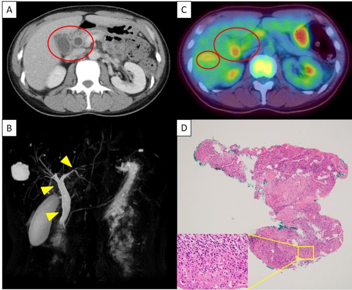
A 50-year-old woman with stage IV lung adenocarcinoma received seven cycles of pembrolizumab as third-line chemotherapy. Following the failure of pembrolizumab, she commenced fourth-line chemotherapy of docetaxel and ramucirumab. The patient complained of epigastric pain and a computed tomography (CT) scan revealed oedema-like thickening of the gallbladder wall, dilation of the bile ducts from the common to the intrahepatic bile ducts, and thickening of the common bile duct wall without any visible obstructions. Accumulation of fluorodeoxyglucose (FDG) in the gallbladder wall and bile duct was also detected with positron emission tomography (PET)-CT. A biopsy of the extrahepatic bile duct showed non-specific inflammation. Antibiotic treatment was not effective and pathogens were not detected. The patient was diagnosed with secondary sclerosing cholangitis (SSC) by pembrolizumab. She received 80 mg/day of prednisolone (PSL); however, SSC recurred with tapering of PSL. SSC then improved with steroid pulse therapy and subsequently 50 mg/day azathioprine and 80 mg/day PSL.
一名50岁的IV期肺腺癌女性接受了七个周期的帕博利珠单抗作为三线化疗。帕博利珠单抗治疗失败后,她开始接受多西他赛和雷莫西尤单抗的四线化疗。患者主诉上腹部疼痛,计算机断层扫描(CT)显示胆囊壁呈水肿样增厚,肝外胆管至肝内胆管扩张,胆总管壁增厚,但未见明显梗阻。正电子发射断层扫描(PET)-CT也检测到胆囊壁和胆管内有氟脱氧葡萄糖(FDG)积聚。肝外胆管活检显示为非特异性炎症。抗生素治疗无效,未检测到病原体。该患者被诊断为帕博利珠单抗所致的继发性硬化性胆管炎(SSC)。她接受了80毫克/天的泼尼松龙(PSL)治疗;然而,随着PSL剂量的逐渐减少,SSC复发。随后,SSC通过类固醇冲击疗法得到改善,随后接受50毫克/天的硫唑嘌呤和80毫克/天的PSL治疗。