• 文献检索
  • 文档翻译
  • 深度研究
  • 学术资讯
  • Suppr Zotero 插件Zotero 插件
  • 邀请有礼
  • 套餐&价格
  • 历史记录
应用&插件
Suppr Zotero 插件Zotero 插件浏览器插件Mac 客户端Windows 客户端微信小程序
定价
高级版会员购买积分包购买API积分包
服务
文献检索文档翻译深度研究API 文档MCP 服务
关于我们
关于 Suppr公司介绍联系我们用户协议隐私条款
关注我们

Suppr 超能文献

核心技术专利:CN118964589B侵权必究
粤ICP备2023148730 号-1Suppr @ 2026

文献检索

告别复杂PubMed语法,用中文像聊天一样搜索,搜遍4000万医学文献。AI智能推荐,让科研检索更轻松。

立即免费搜索

文件翻译

保留排版,准确专业,支持PDF/Word/PPT等文件格式,支持 12+语言互译。

免费翻译文档

深度研究

AI帮你快速写综述,25分钟生成高质量综述,智能提取关键信息,辅助科研写作。

立即免费体验

猪生长和体外成熟卵母细胞的超微结构特征

Ultrastructural Characterization of Porcine Growing and In Vitro Matured Oocytes.

作者信息

Kere Michel, Liu Pan-Chen, Chen Yuh-Kun, Chao Pei-Chi, Tsai Li-Kuang, Yeh Ting-Yu, Siriboon Chawalit, Intawicha Payungsuk, Lo Neng-Wen, Chiang Hsing-I, Fan Yang-Kwang, Ju Jyh-Cherng

机构信息

Department of Animal Science, National Chung Hsing University, 250 Kuo Kuang Road, Taichung 402, Taiwan.

Institute of Rural Development, Nazi Boni University, 01 P.O. Box 1091 Bobo-Dioulasso 01, Burkina Faso.

出版信息

Animals (Basel). 2020 Apr 11;10(4):664. doi: 10.3390/ani10040664.

DOI:10.3390/ani10040664
PMID:32290459
原文链接:https://pmc.ncbi.nlm.nih.gov/articles/PMC7222836/
Abstract

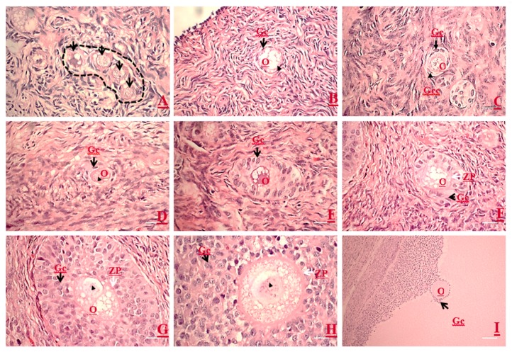
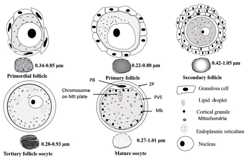
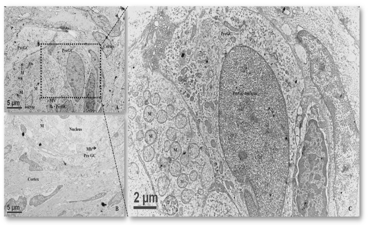
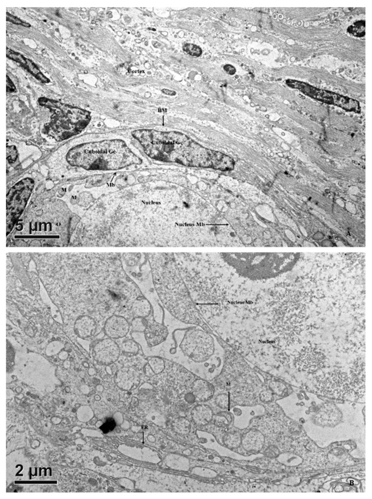
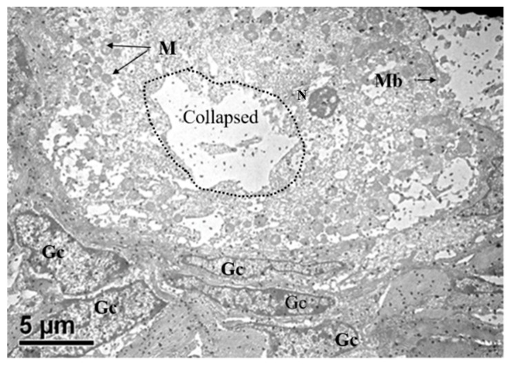
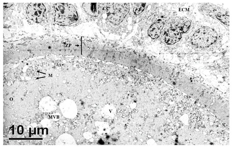
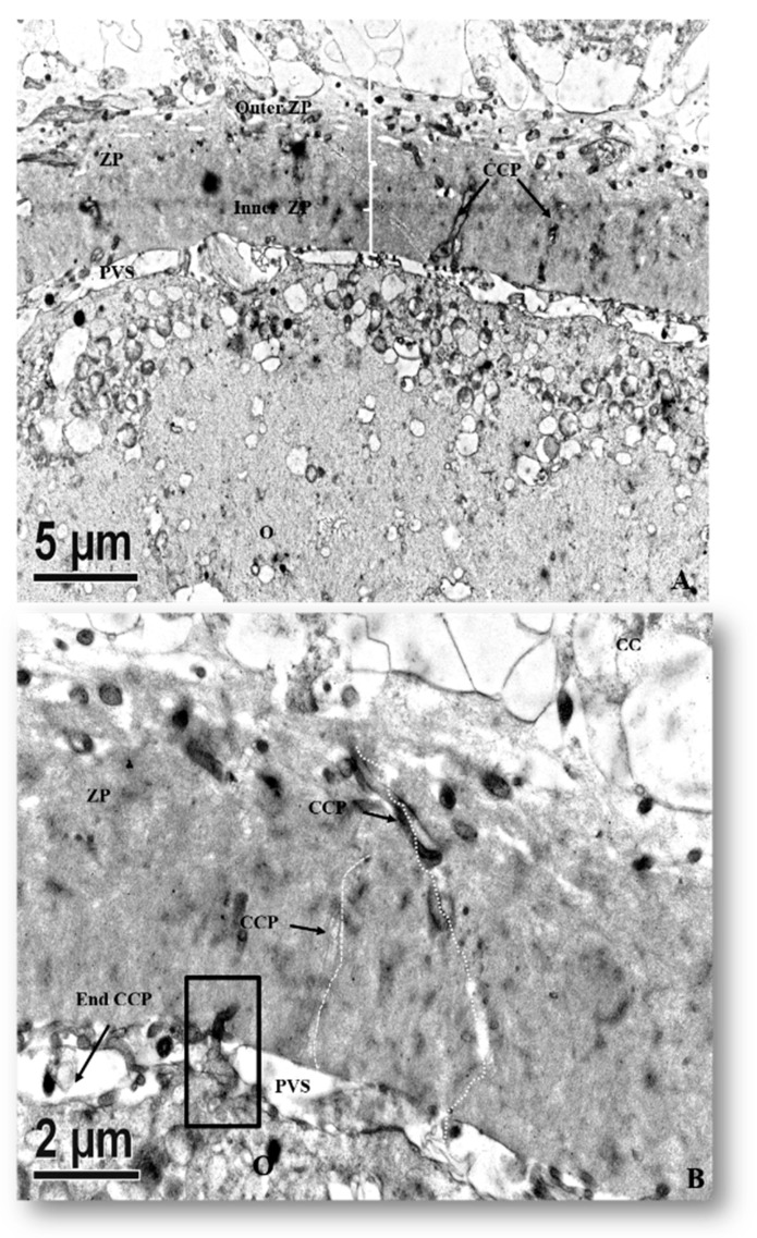
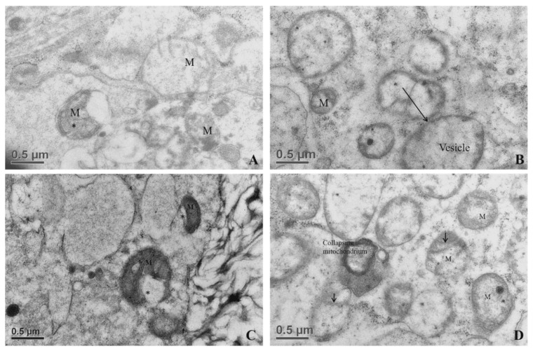
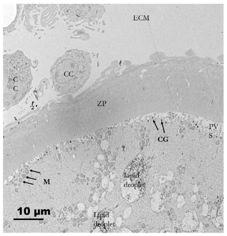
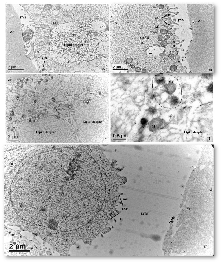
This study aimed to investigate ultrastructural changes of growing porcine oocytes and in vitro maturated oocytes. Light microscopy was used to characterize and localize the primordial, primary, secondary, and tertiary follicles. During oocyte growth and maturation, the morphology of mitochondria was roundish or ovoid in shape depending on the differentiation state, whereas their mean diameters oscillated between 0.5 and 0.7 µm, respectively, from primary and secondary follicles. Hooded mitochondria were found in the growing oocytes of the tertiary follicles. In addition to the pleomorphism of mitochondria, changes in the appearance of lipid droplets were also observed, along with the alignment of a single layer of cortical granules beneath the oolemma. In conclusion, our study is apparently the first report to portray morphological alterations of mitochondria that possess the hooded structure during the growth phase of porcine oocytes. The spatiotemporal and intrinsic changes during oogenesis/folliculogenesis are phenomena at the ultrastructural or subcellular level of porcine oocytes, highlighting an in-depth understanding of oocyte biology and impetus for future studies on practical mitochondrion replacement therapies for oocytes.

摘要

本研究旨在探究生长中的猪卵母细胞和体外成熟卵母细胞的超微结构变化。利用光学显微镜对原始卵泡、初级卵泡、次级卵泡和三级卵泡进行特征描述和定位。在卵母细胞生长和成熟过程中,线粒体的形态根据分化状态呈圆形或椭圆形,而初级卵泡和次级卵泡中线粒体的平均直径分别在0.5至0.7微米之间波动。在三级卵泡的生长中的卵母细胞中发现了带帽线粒体。除了线粒体的多形性外,还观察到脂滴外观的变化,以及卵黄膜下单层皮质颗粒的排列。总之,我们的研究显然是第一份描绘猪卵母细胞生长阶段具有带帽结构的线粒体形态改变的报告。卵子发生/卵泡发生过程中的时空和内在变化是猪卵母细胞超微结构或亚细胞水平的现象,突出了对卵母细胞生物学的深入理解,并为未来卵母细胞实际线粒体替代疗法的研究提供了动力。

https://cdn.ncbi.nlm.nih.gov/pmc/blobs/bdb6/7222836/8051e60f442c/animals-10-00664-g010.jpg
https://cdn.ncbi.nlm.nih.gov/pmc/blobs/bdb6/7222836/e27cd2173397/animals-10-00664-g001.jpg
https://cdn.ncbi.nlm.nih.gov/pmc/blobs/bdb6/7222836/d1e4bda43741/animals-10-00664-g002.jpg
https://cdn.ncbi.nlm.nih.gov/pmc/blobs/bdb6/7222836/0cf3bd73f691/animals-10-00664-g003.jpg
https://cdn.ncbi.nlm.nih.gov/pmc/blobs/bdb6/7222836/47bf241ead92/animals-10-00664-g004.jpg
https://cdn.ncbi.nlm.nih.gov/pmc/blobs/bdb6/7222836/8d5f5cecd056/animals-10-00664-g005.jpg
https://cdn.ncbi.nlm.nih.gov/pmc/blobs/bdb6/7222836/26e8564491f6/animals-10-00664-g006.jpg
https://cdn.ncbi.nlm.nih.gov/pmc/blobs/bdb6/7222836/12130e99beaf/animals-10-00664-g007.jpg
https://cdn.ncbi.nlm.nih.gov/pmc/blobs/bdb6/7222836/dbfbfd4218b4/animals-10-00664-g008.jpg
https://cdn.ncbi.nlm.nih.gov/pmc/blobs/bdb6/7222836/fdb59ec57ccb/animals-10-00664-g009.jpg
https://cdn.ncbi.nlm.nih.gov/pmc/blobs/bdb6/7222836/8051e60f442c/animals-10-00664-g010.jpg

相似文献

1
Ultrastructural Characterization of Porcine Growing and In Vitro Matured Oocytes.猪生长和体外成熟卵母细胞的超微结构特征
Animals (Basel). 2020 Apr 11;10(4):664. doi: 10.3390/ani10040664.
2
Ultrastructural characterization of porcine oocytes and adjacent follicular cells during follicle development: lipid component evolution.猪卵母细胞和卵泡发育过程中相邻卵泡细胞的超微结构特征:脂质成分的演变。
Theriogenology. 2011 Dec;76(9):1647-57. doi: 10.1016/j.theriogenology.2011.06.029. Epub 2011 Aug 10.
3
Oocyte ultrastructure in bovine primordial to early tertiary follicles.牛原始卵泡至早期三级卵泡中的卵母细胞超微结构
Anat Embryol (Berl). 1997 Apr;195(4):327-36. doi: 10.1007/s004290050052.
4
Structural and endocrine aspects of equine oocyte maturation in vivo.马体内卵母细胞成熟的结构和内分泌方面
Mol Reprod Dev. 1995 Sep;42(1):94-105. doi: 10.1002/mrd.1080420113.
5
In vitro maturation is slowed in prepubertal lamb oocytes: ultrastructural evidences.青春期前羔羊卵母细胞的体外成熟减慢:超微结构证据。
Reprod Biol Endocrinol. 2014 Nov 24;12:115. doi: 10.1186/1477-7827-12-115.
6
Ultrastructural changes in oocytes during folliculogenesis in domestic mammals.家畜卵泡发生过程中卵母细胞的超微结构变化
J Ovarian Res. 2014 Oct 30;7:102. doi: 10.1186/s13048-014-0102-6.
7
Oocyte growth and acquisition of meiotic competence.卵母细胞生长与减数分裂能力的获得。
Soc Reprod Fertil Suppl. 2007;63:531-8.
8
From oogonia to mature oocytes: inactivation of the maternal centrosome in humans.从卵原细胞到成熟卵母细胞:人类母体中心体的失活
Microsc Res Tech. 2006 Jun;69(6):396-407. doi: 10.1002/jemt.20299.
9
Oocyte morphology from primordial to early tertiary follicles of yak.牦牛从原始卵泡到早期三级卵泡的卵母细胞形态
Reprod Domest Anim. 2010 Oct;45(5):779-85. doi: 10.1111/j.1439-0531.2009.01347.x.
10
Developmental stages of primary oocytes in turkeys.火鸡初级卵母细胞的发育阶段
Poult Sci. 1996 Dec;75(12):1569-78. doi: 10.3382/ps.0751569.

引用本文的文献

1
Transcriptome Sequencing and Differential Analysis of Ovaries Across Diverse States (Follicular and Non-Follicular Phases).不同状态(卵泡期和非卵泡期)卵巢的转录组测序及差异分析
Animals (Basel). 2025 Aug 20;15(16):2436. doi: 10.3390/ani15162436.
2
Effects of electromagnetic waves on oocyte maturation and embryonic development in pigs.电磁波对猪卵母细胞成熟和胚胎发育的影响。
J Reprod Dev. 2021 Dec 14;67(6):392-401. doi: 10.1262/jrd.2021-074. Epub 2021 Oct 23.

本文引用的文献

1
Ultrastructural changes in oocytes during folliculogenesis in domestic mammals.家畜卵泡发生过程中卵母细胞的超微结构变化
J Ovarian Res. 2014 Oct 30;7:102. doi: 10.1186/s13048-014-0102-6.
2
Pre-spermiogenic initiation of flagellar growth and correlative ultrastructural observations on nuage, nuclear and mitochondrial developmental morphology in the zebrafish Danio rerio.斑马鱼(Danio rerio)精子发生前鞭毛生长的起始以及与生殖质、细胞核和线粒体发育形态相关的超微结构观察
Micron. 2014 Nov;66:1-8. doi: 10.1016/j.micron.2014.03.010. Epub 2014 Mar 31.
3
Vascular endothelial growth factor A improves quality of matured porcine oocytes and developing parthenotes.
血管内皮生长因子A可改善成熟猪卵母细胞和孤雌胚胎的发育质量。
Domest Anim Endocrinol. 2014 Oct;49:60-9. doi: 10.1016/j.domaniend.2014.06.002. Epub 2014 Jun 25.
4
Modifiers of ovarian function in girls and women with classic galactosemia.经典半乳糖血症女性和女童卵巢功能的调节剂。
J Clin Endocrinol Metab. 2013 Jul;98(7):E1257-65. doi: 10.1210/jc.2013-1374. Epub 2013 May 20.
5
Ultrastructure of germ cells, Sertoli cells and mitochondria during spermatogenesis in mature testis of the Chinese Taihang black goats (Capra hircus).中国太行黑山羊(Capra hircus)成熟睾丸精子发生过程中精原细胞、支持细胞和线粒体的超微结构。
Micron. 2013 Jul;50:14-9. doi: 10.1016/j.micron.2013.03.008. Epub 2013 Apr 6.
6
Ascorbic acid improves the developmental competence of porcine oocytes after parthenogenetic activation and somatic cell nuclear transplantation.抗坏血酸可提高猪卵母细胞孤雌激活和体细胞核移植后的发育能力。
J Reprod Dev. 2013;59(1):78-84. doi: 10.1262/jrd.2012-114. Epub 2012 Nov 15.
7
Next generation organelles: structure and role of germ granules in the germline.下一代细胞器:生殖质颗粒在生殖系中的结构和功能。
Mol Reprod Dev. 2013 Aug;80(8):610-23. doi: 10.1002/mrd.22115. Epub 2012 Nov 13.
8
Ultrastructure of human gametes, fertilization and embryos in assisted reproduction: a personal survey.人类配子、受精和胚胎的超微结构在辅助生殖中的研究:个人综述。
Micron. 2013 Jan;44:1-20. doi: 10.1016/j.micron.2012.05.002. Epub 2012 May 18.
9
Nuage proteins: their localization in subcellular structures of spermatogenic cells as revealed by immunoelectron microscopy.云雾蛋白:免疫电镜显示其在精子发生细胞亚细胞结构中的定位。
Histochem Cell Biol. 2012 Jul;138(1):1-11. doi: 10.1007/s00418-012-0962-z. Epub 2012 May 15.
10
Ultrastructural characterization of porcine oocytes and adjacent follicular cells during follicle development: lipid component evolution.猪卵母细胞和卵泡发育过程中相邻卵泡细胞的超微结构特征:脂质成分的演变。
Theriogenology. 2011 Dec;76(9):1647-57. doi: 10.1016/j.theriogenology.2011.06.029. Epub 2011 Aug 10.