• 文献检索
  • 文档翻译
  • 深度研究
  • 学术资讯
  • Suppr Zotero 插件Zotero 插件
  • 邀请有礼
  • 套餐&价格
  • 历史记录
应用&插件
Suppr Zotero 插件Zotero 插件浏览器插件Mac 客户端Windows 客户端微信小程序
定价
高级版会员购买积分包购买API积分包
服务
文献检索文档翻译深度研究API 文档MCP 服务
关于我们
关于 Suppr公司介绍联系我们用户协议隐私条款
关注我们

Suppr 超能文献

核心技术专利:CN118964589B侵权必究
粤ICP备2023148730 号-1Suppr @ 2026

文献检索

告别复杂PubMed语法,用中文像聊天一样搜索,搜遍4000万医学文献。AI智能推荐,让科研检索更轻松。

立即免费搜索

文件翻译

保留排版,准确专业,支持PDF/Word/PPT等文件格式,支持 12+语言互译。

免费翻译文档

深度研究

AI帮你快速写综述,25分钟生成高质量综述,智能提取关键信息,辅助科研写作。

立即免费体验

孤立性胰腺结核的超声成像特征

Ultrasound imaging features of isolated pancreatic tuberculosis.

作者信息

Dong Yi, Jürgensen Christian, Puri Rajesh, D'Onofrio Mirko, Hocke Michael, Wang Wen-Ping, Atkinson Nathan, Sharma Malay, Dietrich Christoph F

机构信息

Department of Ultrasound, Zhongshan Hospital, Fudan University, 200032 Shanghai, China.

Department of Hepatology and Gastroenterology, Charite University, 10117 Berlin, Germany.

出版信息

Endosc Ultrasound. 2018 Mar-Apr;7(2):119-127. doi: 10.4103/2303-9027.210901.

DOI:10.4103/2303-9027.210901
PMID:28721972
原文链接:https://pmc.ncbi.nlm.nih.gov/articles/PMC5914183/
Abstract

BACKGROUND AND OBJECTIVES

Isolated pancreatic tuberculosis (PTB) is extremely rare worldwide. The purpose of this multicenter retrospective study is to analyze imaging features of histologically confirmed isolated PTB in order to determine the diagnostic features of the new methods contrast enhanced ultrasound (CEUS), ultrasound elastography and contrast enhanced endoscopic ultrasound (CE-EUS).

PATIENTS AND METHODS

: We report on a retrospective data collection of 12 cases of PTB confirmed by histology or cytology. All examinations were interpreted by two independent readers in consensus. CEUS, CE-EUS and ultrasound elastography were performed according to the European Federation of Societies for Ultrasound in Medicine and Biology guidelines.

RESULTS

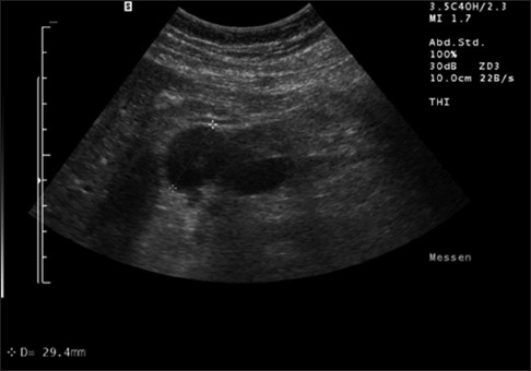
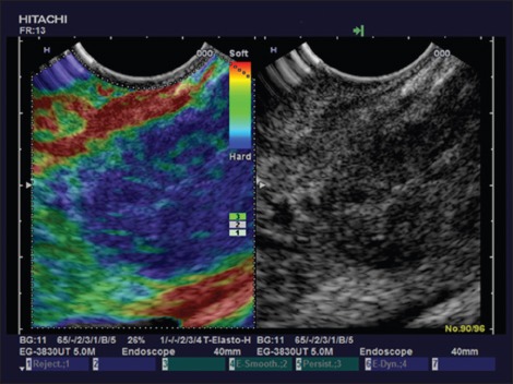
: In PTB patients the common bile duct was never dilated. Multiple retroperitoneal lymph nodes are the second important B-mode ultrasound feature detected in 75% of PTB patients. CE-EUS was performed in three PTB patients demonstrating hyperenhancement. On elastography, all PTB lesions were markedly stiffer than surrounding pancreatic parenchyma.

CONCLUSIONS

: Here we report the first time on CEUS and elastography features of PTB. PTB had some typical imaging features with iso- or hyperenhancement on CE(E) US. PTB is markedly stiffer on elastography. If clinicians are aware of clinical features of PTB and conduct appropriate investigations with multiple modalities including B-mode ultrasound, CEUS, and EUS guided fine needle aspiration, diagnosis of PTB without laparotomy is possible and the disease can be effectively treated with anti-tuberculous drugs.

摘要

背景与目的

孤立性胰腺结核(PTB)在全球极为罕见。这项多中心回顾性研究的目的是分析经组织学确诊的孤立性PTB的影像学特征,以确定新型方法对比增强超声(CEUS)、超声弹性成像和对比增强内镜超声(CE-EUS)的诊断特征。

患者与方法

我们报告了12例经组织学或细胞学确诊的PTB患者的回顾性数据收集情况。所有检查均由两名独立阅片者达成共识后解读。CEUS、CE-EUS和超声弹性成像均按照欧洲医学与生物学超声学会联合会的指南进行。

结果

在PTB患者中,胆总管从未扩张。多个腹膜后淋巴结是在75%的PTB患者中检测到的第二重要的B超特征。对3例PTB患者进行了CE-EUS检查,显示为高增强。在弹性成像中,所有PTB病变均比周围胰腺实质明显更硬。

结论

我们首次报告了PTB的CEUS和弹性成像特征。PTB在CE(E)US上具有一些典型的影像学特征,表现为等增强或高增强。PTB在弹性成像上明显更硬。如果临床医生了解PTB的临床特征,并通过包括B超、CEUS和EUS引导下细针穿刺在内的多种方式进行适当检查,则无需开腹即可诊断PTB,并且该疾病可以用抗结核药物有效治疗。

https://cdn.ncbi.nlm.nih.gov/pmc/blobs/a719/5914183/21afd95ef6c3/EUS-7-119-g006.jpg
https://cdn.ncbi.nlm.nih.gov/pmc/blobs/a719/5914183/fdf33c4b0957/EUS-7-119-g002.jpg
https://cdn.ncbi.nlm.nih.gov/pmc/blobs/a719/5914183/8fa5f6aeccd7/EUS-7-119-g004.jpg
https://cdn.ncbi.nlm.nih.gov/pmc/blobs/a719/5914183/21afd95ef6c3/EUS-7-119-g006.jpg
https://cdn.ncbi.nlm.nih.gov/pmc/blobs/a719/5914183/fdf33c4b0957/EUS-7-119-g002.jpg
https://cdn.ncbi.nlm.nih.gov/pmc/blobs/a719/5914183/8fa5f6aeccd7/EUS-7-119-g004.jpg
https://cdn.ncbi.nlm.nih.gov/pmc/blobs/a719/5914183/21afd95ef6c3/EUS-7-119-g006.jpg

相似文献

1
Ultrasound imaging features of isolated pancreatic tuberculosis.孤立性胰腺结核的超声成像特征
Endosc Ultrasound. 2018 Mar-Apr;7(2):119-127. doi: 10.4103/2303-9027.210901.
2
Autoimmune pancreatitis: Imaging features.自身免疫性胰腺炎:影像学特征。
Endosc Ultrasound. 2018 May-Jun;7(3):196-203. doi: 10.4103/eus.eus_23_17.
3
Direct Comparison of Elastography Endoscopic Ultrasound Fine-Needle Aspiration and B-Mode Endoscopic Ultrasound Fine-Needle Aspiration in Diagnosing Solid Pancreatic Lesions.弹性成像内镜超声细针穿刺与B型内镜超声细针穿刺在诊断胰腺实性病变中的直接比较
Int J Environ Res Public Health. 2022 Jan 24;19(3):1302. doi: 10.3390/ijerph19031302.
4
The Role of Real Time Endoscopic Ultrasound Guided Elastography for Targeting EUS-FNA of Suspicious Pancreatic Masses: A Review of the Literature and A Single Center Experience.实时内镜超声引导弹性成像在可疑胰腺肿块EUS-FNA靶向中的作用:文献综述及单中心经验
JOP. 2016 Sep;17(5):516-524.
5
Endoscopic ultrasound elastography for evaluation of lymph nodes and pancreatic masses: a multicenter study.内镜超声弹性成像用于评估淋巴结和胰腺肿块:一项多中心研究。
World J Gastroenterol. 2009 Apr 7;15(13):1587-93. doi: 10.3748/wjg.15.1587.
6
Endoscopic ultrasound elastography: Current status and future perspectives.内镜超声弹性成像:现状与未来展望。
World J Gastroenterol. 2015 Dec 21;21(47):13212-24. doi: 10.3748/wjg.v21.i47.13212.
7
Endoscopic Ultrasound in the Diagnosis and Staging of Pancreatic Cancer.内镜超声在胰腺癌诊断及分期中的应用
GE Port J Gastroenterol. 2015 Jul 2;22(4):161-171. doi: 10.1016/j.jpge.2015.04.007. eCollection 2015 Jul-Aug.
8
Contrast-enhanced (endoscopic) ultrasound and endoscopic ultrasound elastography in gastrointestinal stromal tumors.对比增强(内镜)超声及内镜超声弹性成像在胃肠道间质瘤中的应用
Endosc Ultrasound. 2017 Jan-Feb;6(1):55-60. doi: 10.4103/2303-9027.200216.
9
Can elastography replace fine needle aspiration?弹性成像能否替代细针抽吸?
Endosc Ultrasound. 2014 Apr;3(2):109-17. doi: 10.4103/2303-9027.123009.
10
New ultrasound techniques for lymph node evaluation.新的超声技术用于淋巴结评估。
World J Gastroenterol. 2013 Aug 14;19(30):4850-60. doi: 10.3748/wjg.v19.i30.4850.

引用本文的文献

1
Ultrasound innovations in abdominal radiology: techniques and clinical applications in pediatric imaging.腹部放射学中的超声创新:儿科影像中的技术与临床应用
Abdom Radiol (NY). 2025 Apr;50(4):1744-1762. doi: 10.1007/s00261-024-04616-x. Epub 2024 Oct 16.
2
Comments and illustrations of the European Federation of Societies for Ultrasound in Medicine contrast-enhanced ultrasound guidelines. Rare pancreatic tumors, imaging features on transabdominal ultrasound and EUS with contrast enhancement: Rare epithelial pancreatic tumors: solid pseudopapillary neoplasm, acinar cell carcinoma, mixed neuroendocrine-non-neuroendocrine neoplasms, some rare subtypes of pancreatic adenocarcinoma and pancreatoblastoma.欧洲医学超声学会对比增强超声指南的评论与说明。罕见胰腺肿瘤,经腹超声和内镜超声造影增强的影像学特征:罕见上皮性胰腺肿瘤:实性假乳头状肿瘤、腺泡细胞癌、神经内分泌-非神经内分泌混合性肿瘤、胰腺腺癌的一些罕见亚型及胰母细胞瘤。
Endosc Ultrasound. 2024 May-Jun;13(3):129-144. doi: 10.1097/eus.0000000000000056. Epub 2024 Apr 30.

本文引用的文献

1
Endobronchial ultrasound elastography.支气管内超声弹性成像。
Endosc Ultrasound. 2016 Jul-Aug;5(4):233-8. doi: 10.4103/2303-9027.187866.
2
EFSUMB Guidelines on Interventional Ultrasound (INVUS), Part V - EUS-Guided Therapeutic Interventions (short version).欧洲超声医学与生物学联合会介入超声指南(INVUS),第五部分 - 超声内镜引导下的治疗性干预(简短版)
Ultraschall Med. 2016 Aug;37(4):412-20. doi: 10.1055/s-0035-1553742. Epub 2016 Aug 4.
3
European Federation of Societies for Ultrasound in Medicine and Biology guidelines 2015 on interventional endoscopic ultrasound.
3
Pancreatic masses clinically diagnosed as tuberculosis: Case reports.临床诊断为结核的胰腺肿块:病例报告
SAGE Open Med Case Rep. 2024 Jun 17;12:2050313X241262192. doi: 10.1177/2050313X241262192. eCollection 2024.
4
Comments and Illustrations of Ultrasound Findings in Extrapulmonary Tuberculosis Manifestations.肺外结核病表现的超声检查结果注释与图示
Diagnostics (Basel). 2024 Mar 27;14(7):706. doi: 10.3390/diagnostics14070706.
5
Misdiagnosis of pancreatic tuberculosis as a pancreatic cystic neoplasm - A case report.胰腺结核误诊为胰腺囊性肿瘤——一例报告
SAGE Open Med Case Rep. 2023 Sep 12;11:2050313X231200289. doi: 10.1177/2050313X231200289. eCollection 2023.
6
Comments on and Illustrations of the EFSUMB CEUS Guidelines: Transabdominal and Endoscopic Ultrasound Features of Intrapancreatic Metastases and the Role of Multiparametric Imaging and EUS-Guided Sampling in Rare Pancreatic Tumors.欧洲超声医学与生物学联合会(EFSUMB)超声造影指南评注与实例:胰腺内转移瘤的经腹及内镜超声特征以及多参数成像和超声内镜引导下采样在罕见胰腺肿瘤中的作用
Cancers (Basel). 2023 Apr 29;15(9):2546. doi: 10.3390/cancers15092546.
7
Cheesy material on macroscopic on-site evaluation after endoscopic ultrasound-guided fine-needle biopsy: Don't miss the tuberculosis.内镜超声引导下细针穿刺活检后宏观现场评估发现的干酪样物质:勿漏诊结核病。
World J Clin Cases. 2023 Apr 6;11(10):2181-2188. doi: 10.12998/wjcc.v11.i10.2181.
8
Current role of endoscopic ultrasound for gastrointestinal and abdominal tuberculosis.内镜超声在胃肠道及腹部结核中的当前作用
JGH Open. 2022 Sep 30;6(11):745-753. doi: 10.1002/jgh3.12823. eCollection 2022 Nov.
9
Exceptional association of hepatic and pancreatic tuberculosis mimicking metastatic pancreatic neoplasia: A case report and review of the literature.酷似转移性胰腺肿瘤的肝结核与胰腺结核罕见关联:一例报告并文献复习
Ann Med Surg (Lond). 2022 May 4;77:103717. doi: 10.1016/j.amsu.2022.103717. eCollection 2022 May.
10
Diagnostic value of endobronchial ultrasound elastography combined with rapid onsite cytological evaluation in endobronchial ultrasound-guided transbronchial needle aspiration.经支气管超声引导针吸活检中,支气管内超声弹性成像联合快速现场细胞学评估的诊断价值。
BMC Pulm Med. 2021 Dec 20;21(1):423. doi: 10.1186/s12890-021-01748-4.
欧洲医学与生物学超声学会联合会2015年介入性内镜超声指南
Endosc Ultrasound. 2016 May-Jun;5(3):143-8. doi: 10.4103/2303-9027.183968.
4
Differential diagnosis of small solid pancreatic lesions.胰腺小实性占位病变的鉴别诊断。
Gastrointest Endosc. 2016 Dec;84(6):933-940. doi: 10.1016/j.gie.2016.04.034. Epub 2016 May 4.
5
EFSUMB Guidelines on Interventional Ultrasound (INVUS), Part IV - EUS-guided interventions: General Aspects and EUS-guided Sampling (Short Version).欧洲超声医学与生物学联合会介入超声指南(INVUS),第四部分 - 超声内镜引导下的介入治疗:一般方面及超声内镜引导下的采样(简短版本)
Ultraschall Med. 2016 Apr;37(2):157-69. doi: 10.1055/s-0035-1553788. Epub 2016 Apr 8.
6
EFSUMB Guidelines on Interventional Ultrasound (INVUS), Part III - Abdominal Treatment Procedures (Short Version).欧洲超声医学与生物学联合会介入超声指南(INVUS),第三部分 - 腹部治疗程序(简短版)
Ultraschall Med. 2016 Feb;37(1):27-45. doi: 10.1055/s-0035-1553965. Epub 2016 Feb 12.
7
Endoscopic ultrasound elastography: Current status and future perspectives.内镜超声弹性成像:现状与未来展望。
World J Gastroenterol. 2015 Dec 21;21(47):13212-24. doi: 10.3748/wjg.v21.i47.13212.
8
EFSUMB Guidelines on Interventional Ultrasound (INVUS), Part III - Abdominal Treatment Procedures (Long Version).欧洲超声医学与生物学联合会介入超声指南(INVUS),第三部分 - 腹部治疗程序(长版)
Ultraschall Med. 2016 Feb;37(1):E1-E32. doi: 10.1055/s-0035-1553917. Epub 2015 Dec 15.
9
EFSUMB Guidelines on Interventional Ultrasound (INVUS), Part II. Diagnostic Ultrasound-Guided Interventional Procedures (Long Version).欧洲超声医学与生物学联合会介入超声指南(INVUS),第二部分。诊断性超声引导介入操作(长篇版)。
Ultraschall Med. 2015 Dec;36(6):E15-35. doi: 10.1055/s-0035-1554036. Epub 2015 Dec 15.
10
EFSUMB Guidelines on Interventional Ultrasound (INVUS), Part II. Diagnostic Ultrasound-Guided Interventional Procedures (Short Version).欧洲超声医学与生物学联合会介入超声指南(INVUS),第二部分。诊断性超声引导介入操作(简短版)。
Ultraschall Med. 2015 Dec;36(6):566-80. doi: 10.1055/s-0035-1566760. Epub 2015 Dec 15.