Department of Neurology, Xuanwu Hospital, Capital Medical University, Beijing, China.
The Beijing Key Laboratory of Neuromodulation, Beijing, China.
Ann Clin Transl Neurol. 2020 May;7(5):653-666. doi: 10.1002/acn3.51033. Epub 2020 Apr 16.
Little is known about the intrinsic electrophysiological properties of hypothalamic hamartoma (HH) in vivo and seizure network since only few cases using stereoelectroencephalography (SEEG) electrodes exploring both cortex and HH have been published. To elucidate these issues, we analyzed simultaneous SEEG recordings in HH and cortex systematically.
We retrospectively investigated data from 15 consecutive patients with SEEG electrodes into the HH for the treatment purpose of radiofrequency thermocoagulation treatment. Additional SEEG electrodes were placed into the cortex in 11 patients to assess extra-HH involvement. Interictal discharges within the HH and anatomo-electroclinical correlations during seizures of each patient were qualitatively and quantitatively analyzed.
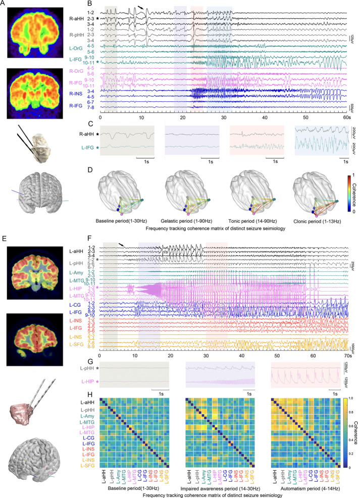
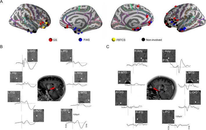
Overall, 77 electrodes with 719 contacts were implanted, and 33 spontaneous seizures were recorded during long-term SEEG monitoring. Interictally, distinct electrophysiological patterns, including isolated intermittent spikes/sharp waves, burst spike and wave trains, paroxysmal fast discharges, periodic discharges, and high-frequency oscillations, were identified within the HH. Notably, synchronized or independent interictal discharges in the cortex were observed. Regarding the ictal discharges, the electrical onset pattern within the HH always started with abrupt giant shifts superimposed on low-voltage fast activity across patients. The gelastic seizure network mainly involved the HH, orbitofrontal areas, and cingulate gyrus. Seizures with automatisms and impaired awareness primarily propagated to mesial temporal lobes. Moreover, independent ictal discharges arising from the mesial temporal lobe were detected in three out of nine patients.
This study comprehensively reveals intrinsic electrophysiological patterns and epileptogenic networks in vivo, providing new insights into the mechanisms underlying cortical and subcortical epileptogenesis.
由于仅少数使用立体脑电图 (SEEG) 电极同时探查皮层和下丘脑错构瘤 (HH) 的病例发表,因此对于 HH 中的内在电生理特性和癫痫网络知之甚少。为了阐明这些问题,我们系统地分析了 HH 和皮层的同时 SEEG 记录。
我们回顾性研究了 15 例连续接受 SEEG 电极植入以进行射频热凝治疗的患者的数据。在 11 例患者中额外放置了 SEEG 电极以评估 HH 以外的累及情况。对每位患者的 HH 内发作间期放电和癫痫发作期间的解剖-电临床相关性进行定性和定量分析。
总共植入了 77 个电极,带有 719 个触点,在长期 SEEG 监测期间记录了 33 次自发性癫痫发作。发作间期,在 HH 内可识别出明显的电生理模式,包括孤立的间歇性棘波/尖波、爆发性棘波和尖波节律、阵发性快速放电、周期性放电和高频振荡。值得注意的是,在皮层中观察到同步或独立的发作间期放电。关于发作期放电,HH 内的电起始模式总是在所有患者中以突然的巨大移位叠加在低电压快速活动上开始。发笑性癫痫网络主要涉及 HH、眶额区和扣带回。伴有自动症和意识障碍的癫痫主要向内侧颞叶传播。此外,在 9 例患者中的 3 例中检测到起源于内侧颞叶的独立发作期放电。
这项研究全面揭示了 HH 内的内在电生理模式和致痫网络,为皮质和皮质下癫痫发生的机制提供了新的见解。