Kronig Otto D M, Kronig Sophia A J, Vrooman Henri A, Veenland Jifke F, Jippes Mariëlle, Boen Terence, Van Adrichem Léon N A
Department of Plastic and Reconstructive Surgery and Hand Surgery, Dutch Craniofacial Centre, Erasmus MC - Sophia Children's Hospital, University Medical Centre Rotterdam, Rotterdam, The Netherlands.
Department of Plastic and Reconstructive Surgery and Hand Surgery, University Medical Centre Utrecht, Heidelberglaan 100, 3584 CX, Utrecht, The Netherlands.
Eur J Pediatr. 2020 Oct;179(10):1569-1577. doi: 10.1007/s00431-020-03643-2. Epub 2020 Apr 17.
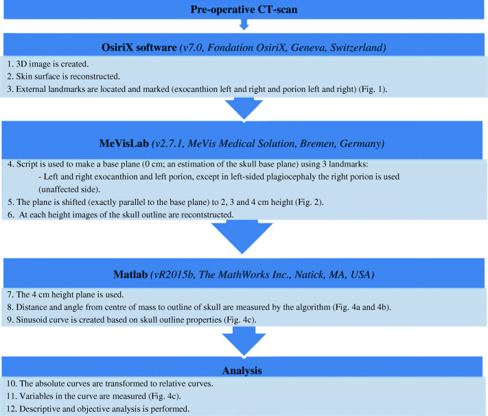
We present a novel technique for classification of skull deformities due to most common craniosynostosis. We included 5 children of every group of the common craniosynostoses (scaphocephaly, brachycephaly, trigonocephaly, and right- and left-sided anterior plagiocephaly) and additionally 5 controls. Our outline-based classification method is described, using the software programs OsiriX, MeVisLab, and Matlab. These programs were used to identify chosen landmarks (porion and exocanthion), create a base plane and a plane at 4 cm, segment outlines, and plot resulting graphs. We measured repeatability and reproducibility, and mean curves of groups were analyzed. All raters achieved excellent intraclass correlation scores (0.994-1.000) and interclass correlation scores (0.989-1.000) for identifying the external landmarks. Controls, scaphocephaly, trigonocephaly, and brachycephaly all have the peak of the forehead in the middle of the curve (180°). In contrary, in anterior plagiocephaly, the peak is shifted (to the left of graph in right-sided and vice versa). Additionally, controls, scaphocephaly, and trigonocephaly have a high peak of the forehead; scaphocephaly has the lowest troughs; in brachycephaly, the width/frontal peak ratio has the highest value with a low frontal peak.Conclusion: We introduced a preliminary study showing an objective and reproducible methodology using CT scans for the analysis of craniosynostosis and potential application of our method to 3D photogrammetry. What is Known: • Diagnosis of craniosynostosis is relatively simple; however, classification of craniosynostosis is difficult and current techniques are not widely applicable. What is New: • We introduce a novel technique for classification of skull deformities due to craniosynostosis, an objective and reproducible methodology using CT scans resulting in characteristic curves. The method is applicable to all 3D-surface rendering techniques. • Using external landmarks and curve analysis, specific and characteristic curves for every type of craniosynostosis related to the specific skull deformities are found.
我们提出了一种用于对最常见的颅缝早闭所致颅骨畸形进行分类的新技术。我们纳入了每组常见颅缝早闭(舟状头、短头、三角头以及右侧和左侧前斜头畸形)的5名儿童,并另外纳入了5名对照。我们描述了基于轮廓的分类方法,使用了OsiriX、MeVisLab和Matlab软件程序。这些程序用于识别选定的标志点(耳点和外眦点),创建一个基准平面和一个4厘米处的平面,分割轮廓,并绘制所得图形。我们测量了重复性和再现性,并分析了各组的平均曲线。所有评估者在识别外部标志点时均获得了出色的组内相关系数得分(0.994 - 1.000)和组间相关系数得分(0.989 - 1.000)。对照组、舟状头、三角头和短头畸形在曲线中间(180°)均有额头峰值。相反,在前斜头畸形中,峰值发生偏移(右侧的在图形左侧,反之亦然)。此外,对照组、舟状头和三角头有较高的额头峰值;舟状头有最低的波谷;在短头畸形中,宽度/额部峰值比具有最高值且额部峰值较低。结论:我们开展了一项初步研究,展示了一种使用CT扫描分析颅缝早闭的客观且可重复的方法,以及我们的方法在三维摄影测量中的潜在应用。已知信息:• 颅缝早闭的诊断相对简单;然而,颅缝早闭的分类困难且当前技术应用不广泛。新内容:• 我们引入了一种用于对颅缝早闭所致颅骨畸形进行分类的新技术,一种使用CT扫描的客观且可重复的方法,可得出特征曲线。该方法适用于所有三维表面渲染技术。• 通过使用外部标志点和曲线分析,发现了与特定颅骨畸形相关的每种颅缝早闭类型的特定且特征性曲线。