Dai P L, Du X S, Hou Y, Li L, Xia Y X, Wang L, Chen H X, Chang L, Li W H
Radiotherapy Department, The Third Affiliated Hospital of Kunming Medical University, Kunming, Yunnan 650100, People's Republic of China.
Kunming Medical University, Kunming, Yunnan 650100, People's Republic of China.
Cancer Manag Res. 2020 Apr 2;12:2437-2447. doi: 10.2147/CMAR.S219967. eCollection 2020.
The biological changes after irradiation in lung cancer cells are important to reduce recurrence and metastasis of lung cancer. To optimize radiotherapy of lung adenocarcinoma, our study systematically explored the mechanisms of biological behaviors in residual A549 and XWLC-05 cells after irradiation.
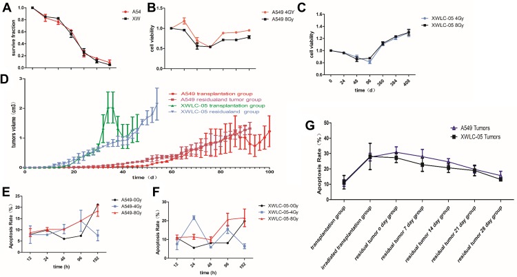
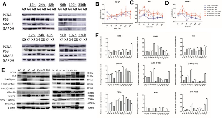
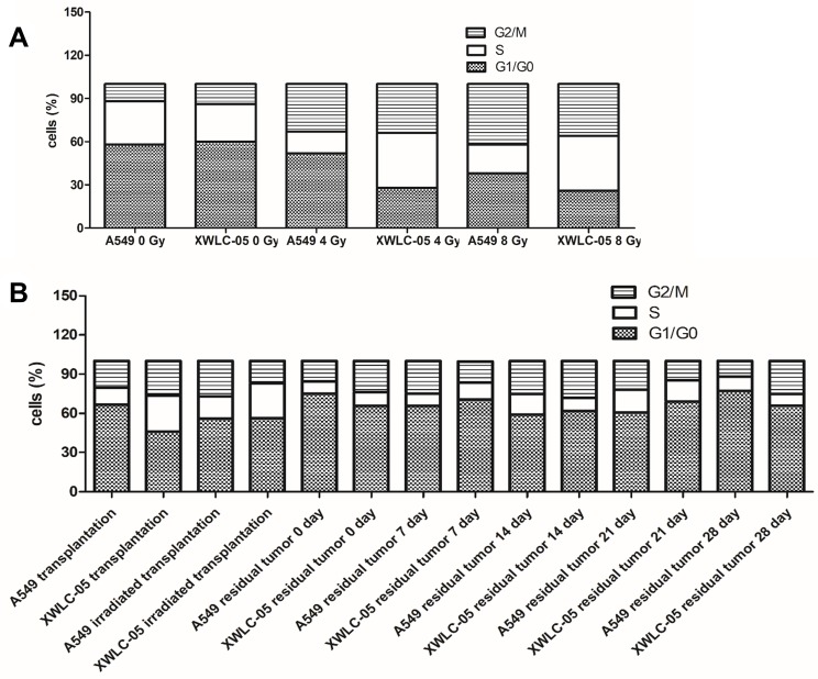
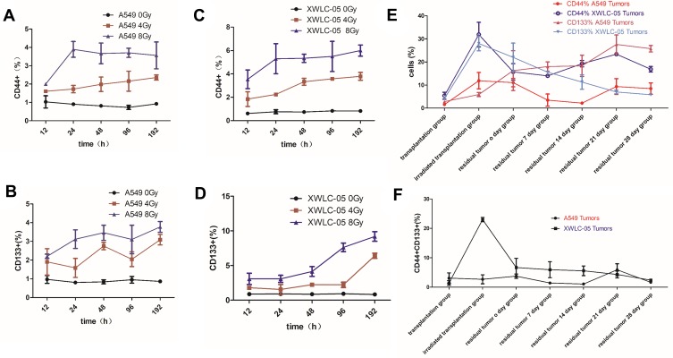
Colony formation assay, cell proliferation assay, cell migration assay, flow cytometry, BALB/C-nu mice xenograft models and Western blot of pan-AKT, p-Akt380, p-Akt473, PCNA, DNA-PKCS, KU70, KU80, CD133, CD144, MMP2 and P53 were used in our study to assess biological changes after irradiation with 0, 4 and 8 Gy at 0-336 hr after irradiation in vitro and 20 Gy at transplantation group, irradiated transplantation group, residual tumor 0, 7, 14, 21, and 28 days groups in vivo.
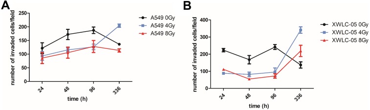
The ability of cell proliferation and radiosensitivity of residual XWLC-05 cells was better than A549 cells after radiation in vivo and in vitro. MMP-2 has statistical differences in vitro and in vivo and increased with the migratory ability of cells in vitro. PCNA and P53 have statistical differences in XWLC-05 and A549 cells and the changes of them are similar to the proliferation of residual cells within first 336 hr after irradiation in vitro. Pan-AKT increased after irradiation, and residual tumor 21-day group (1.5722) has statistic differences between transplantation group (0.9763, p=0.018) and irradiated transplantation group (0.8455, p=0.006) in vivo. Pan-AKT rose to highest when 21-day after residual tumor reach to 0.5 mm. MMP2 has statistical differences between transplantation group (0.4619) and residual tumor 14-day group (0.8729, p=0.043). P53 has statistical differences between residual tumor 7-day group (0.6184) and residual tumor 28 days group (1.0394, p=0.007). DNA-PKCS has statistical differences between residual tumor 28 days group (1.1769) and transplantation group (0.2483, p=0.010), irradiated transplantation group (0.1983, p=0.002) and residual tumor 21 days group (0.2017, p=0.003), residual tumor 0 days group (0.5992) and irradiated transplantation group (0.1983, p=0.027) and residual tumor 21 days group (0.2017, p=0.002). KU80 and KU70 have no statistical differences at any time point.
Different proteins regulated apoptosis, proliferation and metastasis of lung adenocarcinoma after radiotherapy at different times. MMP-2 might regulate metastasis ability of XWLC-05 and A549 cells in vitro and in vivo. PCNA and P53 may play important roles in proliferation of vitro XWLC-05 and A549 cells within first 336 hr after irradiation in vitro. After that, P53 may through PI3K/AKT pathway regulate cell proliferation after irradiation in vitro. DNA-PKCS may play a more important role in DNA damage repair than KU70 and KU80 after 336 hr in vitro because it rapidly rose than KU70 and KU80 after irradiation. Different cells have different time rhythm in apoptosis, proliferation and metastasis after radiotherapy. Time rhythm of cells after irradiation should be delivered and more attention should be paid to resist cancer cell proliferation and metastasis.
肺癌细胞受照射后的生物学变化对于降低肺癌的复发和转移至关重要。为优化肺腺癌的放射治疗,我们的研究系统地探索了照射后残留A549和XWLC - 05细胞生物学行为的机制。
我们的研究使用集落形成试验、细胞增殖试验、细胞迁移试验、流式细胞术、BALB/C - nu小鼠异种移植模型以及针对泛AKT、p - Akt380、p - Akt473、PCNA、DNA - PKCS、KU70、KU80、CD133、CD144、MMP2和P53的蛋白质印迹法,以评估体外照射0、4和8 Gy后0 - 336小时以及体内移植组、照射移植组、残留肿瘤0、7、14、21和28天组照射后的生物学变化。
体内和体外照射后,残留XWLC - 05细胞的增殖能力和放射敏感性均优于A549细胞。MMP - 2在体外和体内均有统计学差异,且随体外细胞迁移能力增加。PCNA和P53在XWLC - 05和A549细胞中有统计学差异,其变化与体外照射后前336小时内残留细胞的增殖情况相似。照射后泛AKT增加,体内残留肿瘤21天组(1.5722)与移植组(0.9763,p = 0.018)和照射移植组(0.8455,p = 0.006)之间存在统计学差异。残留肿瘤达到0.5 mm后21天时泛AKT升至最高。MMP2在移植组(0.4619)和残留肿瘤14天组(0.8729,p = 0.043)之间有统计学差异。P53在残留肿瘤7天组(0.6184)和残留肿瘤28天组(1.0394,p = 0.007)之间有统计学差异。DNA - PKCS在残留肿瘤28天组(1.1769)与移植组(0.2483,p = 0.010)、照射移植组(0.1983,p = 0.002)和残留肿瘤21天组(0.2017,p = 0.003)、残留肿瘤0天组(0.5992)与照射移植组(0.1983,p = 0.027)和残留肿瘤21天组(0.2017,p = 0.002)之间有统计学差异。KU80和KU70在任何时间点均无统计学差异。
不同蛋白质在放疗后的不同时间调节肺腺癌的凋亡、增殖和转移。MMP - 2可能在体外和体内调节XWLC - 05和A549细胞的转移能力。PCNA和P53可能在体外照射后前336小时内对体外XWLC - 05和A549细胞的增殖起重要作用。此后,P53可能通过PI3K/AKT途径调节体外照射后的细胞增殖。体外336小时后,DNA - PKCS在DNA损伤修复中可能比KU70和KU80发挥更重要作用,因为照射后它比KU70和KU80迅速升高。不同细胞在放疗后的凋亡、增殖和转移具有不同的时间节律。应重视照射后细胞的时间节律,更加关注抑制癌细胞的增殖和转移。