Department of Basic Medical Sciences, Faculty of Medicine, The Hashemite University, Zarqa 13115, Jordan.
Department of Pharmacology and Toxicology, College of Pharmacy, King Saud University, Riyadh 11451, Saudi Arabia.
Medicina (Kaunas). 2022 Jul 2;58(7):889. doi: 10.3390/medicina58070889.
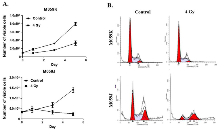
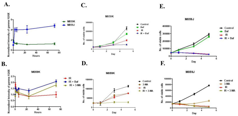
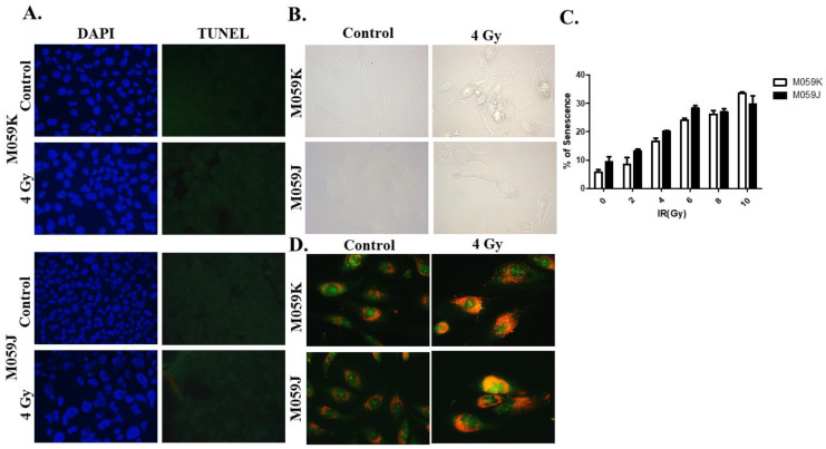
: The development of radioresistance is a fundamental barrier to successful glioblastoma therapy. Autophagy is thought to play a role in facilitating the DNA repair of DNA damage foci in radiation-exposed tumor cells, thus, potentially contributing to their restoration of proliferative capacity and development of resistance in vitro. However, the effect of autophagy inhibitors on DNA damage repair is not fully clear and requires further investigation. : In this work, we utilized M059K (DNA-PKcs proficient) and M059J (DNA-PKcs deficient) glioma cell lines to investigate the role of autophagy inhibitors in the DNA repair of radiation-induced DNA damage. Cell viability following radiation was determined by trypan blue exclusion in both cell lines. Cell death and autophagy assays were performed to evaluate radiation-induced cell stress responses. DNA damage was measured as based on the intensity of phosphorylated γ-H2AX, a DNA double-stranded breaks (DSBs) marker, in the presence or absence of autophagy inhibitors. : The cell viability assay showed that M059J cells were more sensitive to the same dose of radiation (4 Gy) than M059K cells. This observation was accompanied by an elevation in γ-H2AX formation in M059J but not in M059K cells. In addition, the DAPI/TUNEL and Senescence-associated β-galactosidase (SA-β-gal) staining assays did not reveal significant differences in apoptosis and/or senescence induction in response to radiation, respectively, in either cell line. However, acridine orange staining demonstrated clear promotion of acidic vesicular organelles (AVOs) in both cell lines in response to 4 Gy radiation. Moreover, DNA damage marker levels were found to be elevated 72 h post-radiation when autophagy was inhibited by the lysosomotropic agent bafilomycin A1 (BafA1) or the PI3K inhibitor 3-methyl adenine (3-MA) in M059K cells. : The extent of the DNA damage response remained high in the DNA-PKcs deficient cells following exposure to radiation, indicating their inability to repair the newly formed DNA-DSBs. On the other hand, radioresistant M059K cells showed more DNA damage response only when autophagy inhibitors were used with radiation, suggesting that the combination of autophagy inhibitors with radiation may interfere with DNA repair efficiency.
:放射抵抗的发展是胶质母细胞瘤治疗成功的一个基本障碍。自噬被认为在促进放射暴露的肿瘤细胞中 DNA 损伤焦点的 DNA 修复中发挥作用,从而可能有助于它们恢复增殖能力并在体外产生耐药性。然而,自噬抑制剂对 DNA 损伤修复的影响尚不完全清楚,需要进一步研究。:在这项工作中,我们利用 M059K(DNA-PKcs 功能正常)和 M059J(DNA-PKcs 缺陷)神经胶质瘤细胞系来研究自噬抑制剂在放射诱导的 DNA 损伤的 DNA 修复中的作用。在这两种细胞系中,通过台盼蓝排除法测定放射后的细胞活力。进行细胞死亡和自噬测定以评估放射诱导的细胞应激反应。根据存在或不存在自噬抑制剂时磷酸化 γ-H2AX 的强度来测量 DNA 损伤,γ-H2AX 是 DNA 双链断裂 (DSBs) 的标志物。:细胞活力测定表明,M059J 细胞对相同剂量(4 Gy)的辐射比 M059K 细胞更敏感。这一观察结果伴随着 M059J 细胞中 γ-H2AX 形成的升高,但 M059K 细胞中没有。此外,DAPI/TUNEL 和衰老相关 β-半乳糖苷酶 (SA-β-gal) 染色测定分别未显示出两种细胞系中对辐射的凋亡和/或衰老诱导的显著差异。然而,吖啶橙染色显示,在两种细胞系中,在接受 4 Gy 辐射后,均明显促进酸性囊泡细胞器 (AVO) 的形成。此外,当用溶酶体靶向剂巴弗洛霉素 A1 (BafA1) 或 PI3K 抑制剂 3-甲基腺嘌呤 (3-MA) 抑制自噬时,在 M059K 细胞中,72 小时后放射后 DNA 损伤标志物水平升高。:在暴露于辐射后,DNA-PKcs 缺陷细胞中的 DNA 损伤反应程度仍然很高,表明它们无法修复新形成的 DNA-DSBs。另一方面,耐辐射的 M059K 细胞仅在使用自噬抑制剂与辐射联合使用时才显示出更高的 DNA 损伤反应,这表明自噬抑制剂与辐射的联合使用可能会干扰 DNA 修复效率。