Liu Fulin, Rouault Christine, Guesnon Mickael, Zhu Wencan, Clément Karine, Degrelle Séverine A, Fournier Thierry
Université de Paris, INSERM, UMR-S1139 "Pathophysiology & Pharmacotoxicology of the Human Placenta, Pre & Postnatal Microbiota" (3PHM), Paris F-75006, France.
Fondation PremUp, Paris F-75006, France.
PPAR Res. 2020 Apr 1;2020:9210748. doi: 10.1155/2020/9210748. eCollection 2020.
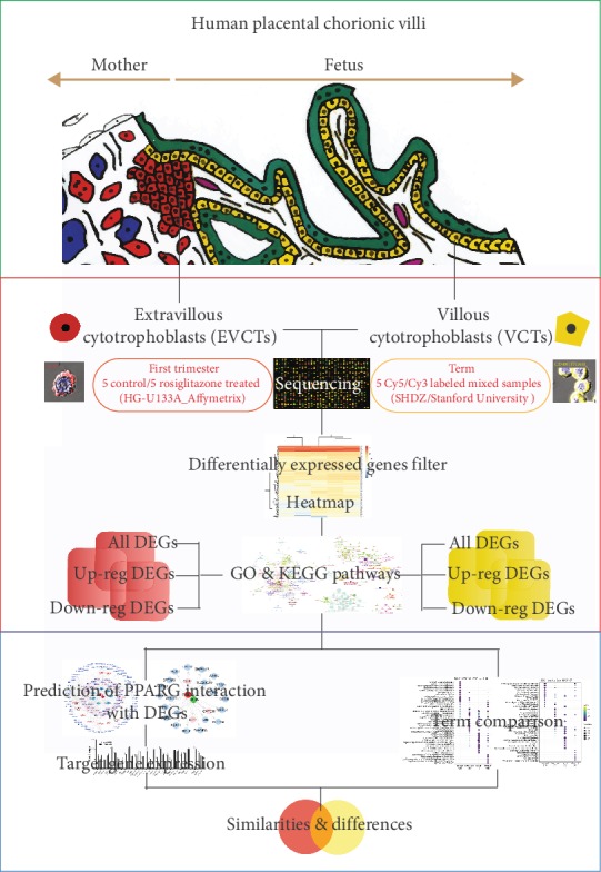
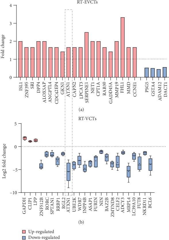
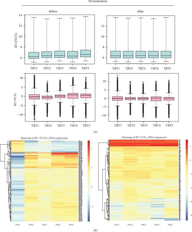
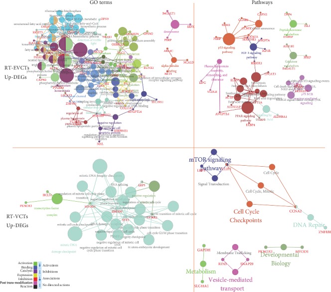
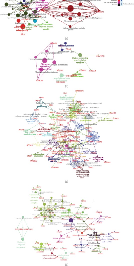
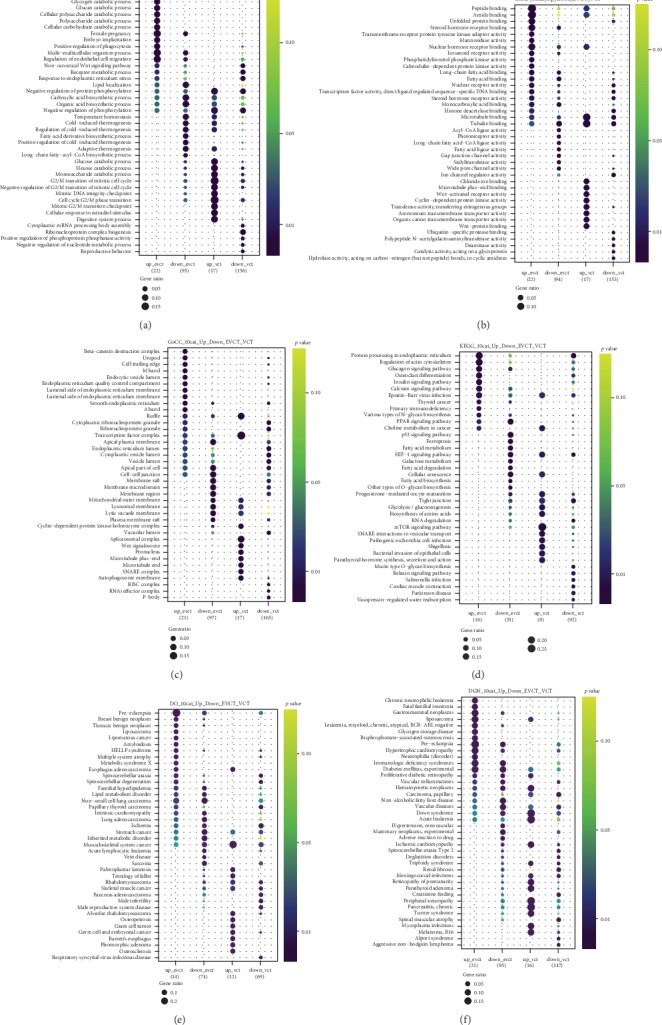
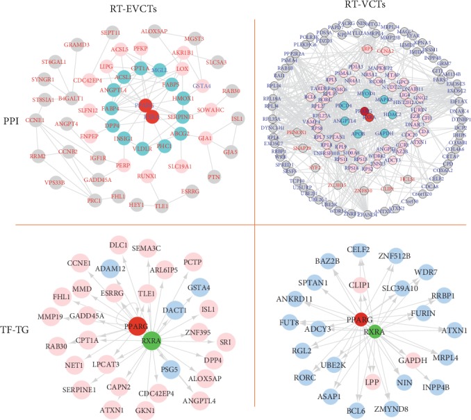
Trophoblasts, as the cells that make up the main part of the placenta, undergo cell differentiation processes such as invasion, migration, and fusion. Abnormalities in these processes can lead to a series of gestational diseases whose underlying mechanisms are still unclear. One protein that has proven to be essential in placentation is the peroxisome proliferator-activated receptor (PPAR), which is expressed in the nuclei of extravillous cytotrophoblasts (EVCTs) in the first trimester and villous cytotrophoblasts (VCTs) throughout pregnancy. Here, we aimed to explore the genome-wide effects of PPAR on EVCTs and VCTs via treatment with the PPAR-agonist rosiglitazone. EVCTs and VCTs were purified from human chorionic villi, cultured , and treated with rosiglitazone. The transcriptomes of both types of cells were then quantified using microarray profiling. Differentially expressed genes (DEGs) were filtered and submitted for gene ontology (GO) annotation and pathway analysis with ClueGO. The online tool STRING was used to predict PPAR and DEG protein interactions, while iRegulon was used to predict the binding sites for PPAR and DEG promoters. GO and pathway terms were compared between EVCTs and VCTs with clusterProfiler. Visualizations were prepared in Cytoscape. From our microarray data, 139 DEGs were detected in rosiglitazone-treated EVCTs (RT-EVCTs) and 197 DEGs in rosiglitazone-treated VCTs (RT-VCTs). Downstream annotation analysis revealed the similarities and differences between RT-EVCTs and RT-VCTs with respect to the biological processes, molecular functions, cellular components, and KEGG pathways affected by the treatment, as well as predicted binding sites for both protein-protein interactions and transcription factor-target gene interactions. These results provide a broad perspective of PPAR-activated processes in trophoblasts; further analysis of the transcriptomic signatures of RT-EVCTs and RT-VCTs should open new avenues for future research and contribute to the discovery of possible drug-targeted genes or pathways in the human placenta.
滋养层细胞作为构成胎盘主要部分的细胞,会经历侵袭、迁移和融合等细胞分化过程。这些过程中的异常会导致一系列妊娠疾病,但其潜在机制仍不清楚。一种在胎盘形成过程中已被证明至关重要的蛋白质是过氧化物酶体增殖物激活受体(PPAR),它在孕早期的绒毛外细胞滋养层(EVCTs)细胞核中表达,并在整个孕期的绒毛细胞滋养层(VCTs)中表达。在此,我们旨在通过用PPAR激动剂罗格列酮处理来探索PPAR对EVCTs和VCTs的全基因组影响。从人绒毛膜绒毛中纯化出EVCTs和VCTs,进行培养并用罗格列酮处理。然后使用微阵列分析对两种类型细胞的转录组进行定量。筛选出差异表达基因(DEGs),并使用ClueGO进行基因本体(GO)注释和通路分析。在线工具STRING用于预测PPAR与DEG的蛋白质相互作用,而iRegulon用于预测PPAR与DEG启动子的结合位点。使用clusterProfiler比较EVCTs和VCTs之间的GO和通路术语。在Cytoscape中进行可视化。从我们的微阵列数据中,在罗格列酮处理的EVCTs(RT-EVCTs)中检测到139个DEGs,在罗格列酮处理的VCTs(RT-VCTs)中检测到197个DEGs。下游注释分析揭示了RT-EVCTs和RT-VCTs在受处理影响的生物学过程、分子功能、细胞成分和KEGG通路方面的异同,以及蛋白质-蛋白质相互作用和转录因子-靶基因相互作用的预测结合位点。这些结果提供了滋养层细胞中PPAR激活过程的广泛视角;对RT-EVCTs和RT-VCTs转录组特征的进一步分析应为未来研究开辟新途径,并有助于发现人胎盘中可能的药物靶向基因或通路。