Islam Md Tauhidul, Tasciotti Ennio, Righetti Raffaella
1Department of Electrical and Computer EngineeringTexas A&M UniversityCollege StationTX77843USA.
2Center of Biomimetic MedicineHouston Methodist Research InstituteHoustonTX77030USA.
IEEE J Transl Eng Health Med. 2019 Sep 13;7:4300209. doi: 10.1109/JTEHM.2019.2932059. eCollection 2019.
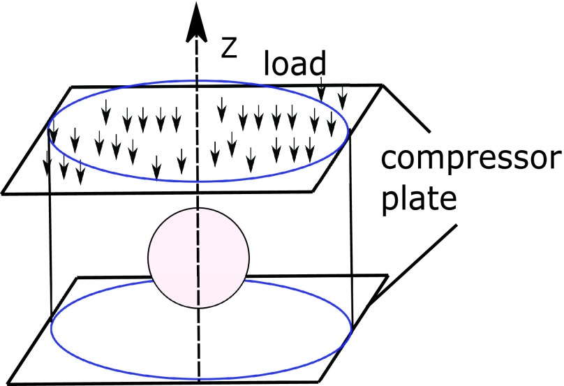
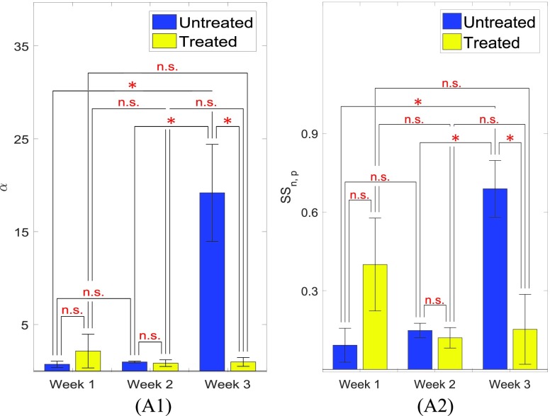
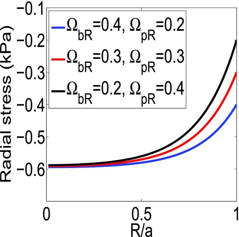
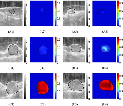
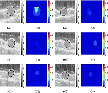
The solid stress (SSg) that develops inside a cancer is an important marker of cancer's growth, invasion and metastasis. Currently, there are no non-invasive methods to image SSg inside tumors. In this paper, we develop a new, non-invasive and cost-effective imaging method to assess SSg inside tumors that uses ultrasound poroelastography. Center to the proposed method is a novel analytical model, which demonstrates that SSg and the compression-induced stress (SSc) that generates inside the cancer in a poroelastography experiment have the same spatial distribution. To show the clinical feasibility of the proposed technique, we imaged and analyzed the normalized SSg inside treated and untreated human breast cancers in a small animal model. Given the clinical significance of assessing SSg in cancers and the advantages of the proposed ultrasonic methods, our technique could have a great impact on cancer diagnosis, prognosis and treatment methods.
癌症内部产生的固体应力(SSg)是癌症生长、侵袭和转移的重要标志物。目前,尚无用于对肿瘤内部的SSg进行成像的非侵入性方法。在本文中,我们开发了一种新的、非侵入性且具有成本效益的成像方法,用于使用超声孔隙弹性成像评估肿瘤内部的SSg。所提出方法的核心是一个新颖的分析模型,该模型表明在孔隙弹性成像实验中,SSg与癌症内部产生的压缩诱导应力(SSc)具有相同的空间分布。为了展示所提出技术的临床可行性,我们在一个小动物模型中对经治疗和未经治疗的人类乳腺癌内部的归一化SSg进行了成像和分析。鉴于评估癌症中SSg的临床意义以及所提出超声方法的优势,我们的技术可能会对癌症诊断、预后和治疗方法产生重大影响。