Chen Pengyuan, Wang Jiaqiang, Wang Xingye, Chen Xiaolin, Li Chunling, Tan Taichang
Sichuan Academy of Medical Sciences & Sichuan Provincial People's Hospital, Chengdu 610072, China.
Department of Laboratory Medicine, Sichuan Provincial People's Hospital, University of Electronic Science and Technology of China, Chengdu 611731, China.
Ann Transl Med. 2020 Mar;8(5):239. doi: 10.21037/atm.2020.03.46.
Maternal embryonic leucine zipper kinase (MELK) is an atypical member of the snf1/AMPK family of serine-threonine kinases, involved in diverse physiological and pathological processes, including cell proliferation, apoptosis, embryogenesis, cancer treatment resistance, and RNA processing. is highly expressed in human cancers and is associated with more aggressive forms of astrocytoma, glioblastoma, breast cancer, and melanoma to date, no information about porcine MELK (pMELK) has been reported.
In this study, the p coding sequence was cloned from swine spleen and characterized. We also quantitatively determined the expression of in 11 tissues isolated from a piglet and determined its subcellular localization when expressed in swine umbilical vein endothelial cells (SUVEC) as a fusion protein. Moreover, we report the functional characterization of pMELK protein concerning its role in apoptosis.
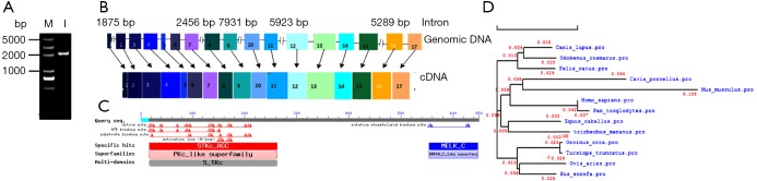
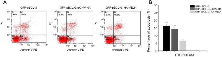
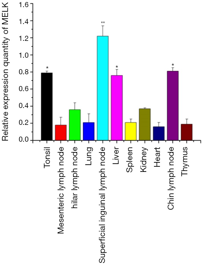
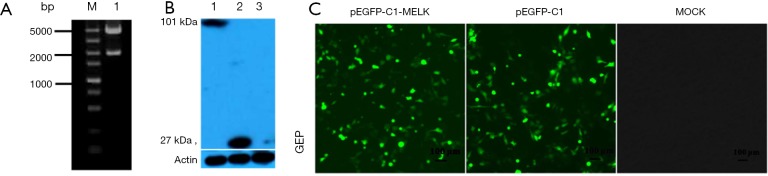
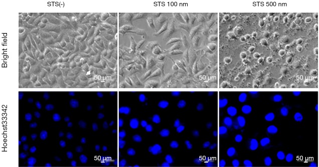
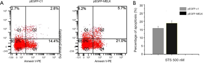
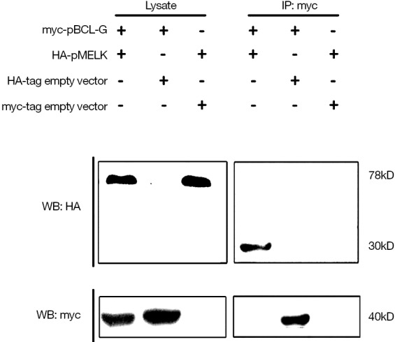
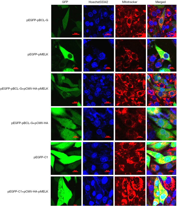
Sequencing analysis showed that full-length of pMELK is 2,072 bp with 17 exons, encoding 655 amino acids, including an S-TKc conserved domain. Comparison of pMELK with ten other mammalian species of their orthologous sequences showed >91% homology and an evolutionary distance <0.05, demonstrating that MELK is highly conserved in evolution. Relative quantification of MELK expression in 11 tissue samples isolated from 30-day-old piglets showed MELK expression in all tested organs and the highest expression in the superficial inguinal lymph node. Constructed a plasmid named pEGFP-MELK, and the fusion protein GFP-MELK was successfully expressed in SUVECs. Fluorescence microscopy revealed the subcellular distribution of the fusion protein GFP-MELK was limited to the cytoplasm. About function, Flow cytometry analysis showed that overexpression of GFP-pMELK in SUVEC cells enhances staurosporine (STS)-induced apoptosis, but not significantly different. The pMELK protein also was found to interact with porcine BCL-G and transient transfection of the recombinant plasmid pCMV-HA-pMELK into SUVEC cells stably expressing GFP-pBCL-G protein inhibited pBCL-G -induced apoptosis significantly.
The present study provided useful information on pMELK basic details and function in apoptosis offer a potential new molecular model for disease interventions and disease related to human MELK and BCL-G.
母体胚胎亮氨酸拉链激酶(MELK)是丝氨酸 - 苏氨酸激酶snf1/AMPK家族的非典型成员,参与多种生理和病理过程,包括细胞增殖、凋亡、胚胎发生、癌症治疗抗性和RNA加工。它在人类癌症中高度表达,并且与更具侵袭性的星形细胞瘤、胶质母细胞瘤、乳腺癌和黑色素瘤相关。迄今为止,尚未有关于猪MELK(pMELK)的信息报道。
在本研究中,从猪脾脏中克隆并鉴定了pMELK的编码序列。我们还定量测定了从一头仔猪分离的11种组织中pMELK的表达,并确定了其在猪脐静脉内皮细胞(SUVEC)中作为融合蛋白表达时的亚细胞定位。此外,我们报告了pMELK蛋白在细胞凋亡中作用的功能特性。
测序分析表明,pMELK全长2072 bp,含17个外显子,编码655个氨基酸,包括一个S-TKc保守结构域。将pMELK与其他十种哺乳动物的直系同源序列进行比较,显示同源性>91%,进化距离<0.05,表明MELK在进化中高度保守。对从30日龄仔猪分离的11个组织样本中MELK表达的相对定量分析表明,MELK在所有测试器官中均有表达,在腹股沟浅淋巴结中表达最高。构建了名为pEGFP-MELK的质粒,融合蛋白GFP-MELK在SUVEC细胞中成功表达。荧光显微镜显示融合蛋白GFP-MELK的亚细胞分布局限于细胞质。关于功能,流式细胞术分析表明,在SUVEC细胞中过表达GFP-pMELK可增强星形孢菌素(STS)诱导的细胞凋亡,但差异不显著。还发现pMELK蛋白与猪BCL-G相互作用,将重组质粒pCMV-HA-pMELK瞬时转染到稳定表达GFP-pBCL-G蛋白的SUVEC细胞中可显著抑制pBCL-G诱导的细胞凋亡。
本研究提供了关于pMELK基本细节及其在细胞凋亡中功能的有用信息,为与人类MELK和BCL-G相关的疾病干预提供了一个潜在的新分子模型。