da Silva Luiza de Campos Moreira, de Oliveira Julia Teixeira, Tochetto Sandra, de Oliveira Claudia Pinto Marques Souza, Sigrist Rosa, Chammas Maria Cristina
Faculdade de Medicina da Universidade de São Paulo (FMUSP), São Paulo, SP, Brazil.
Radiol Bras. 2020 Jan-Feb;53(1):47-55. doi: 10.1590/0100-3984.2019.0028.
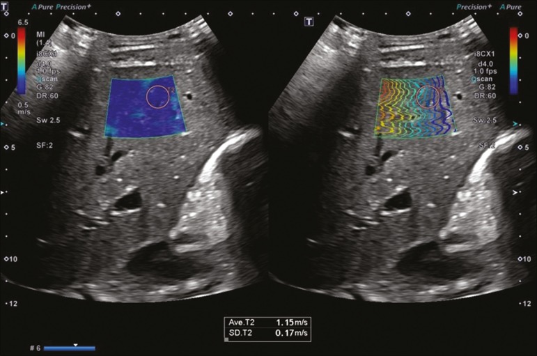
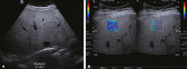
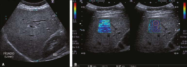
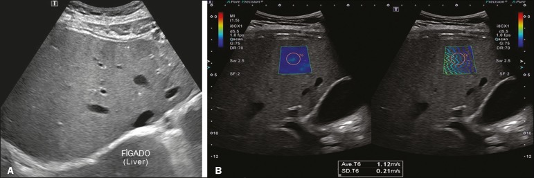
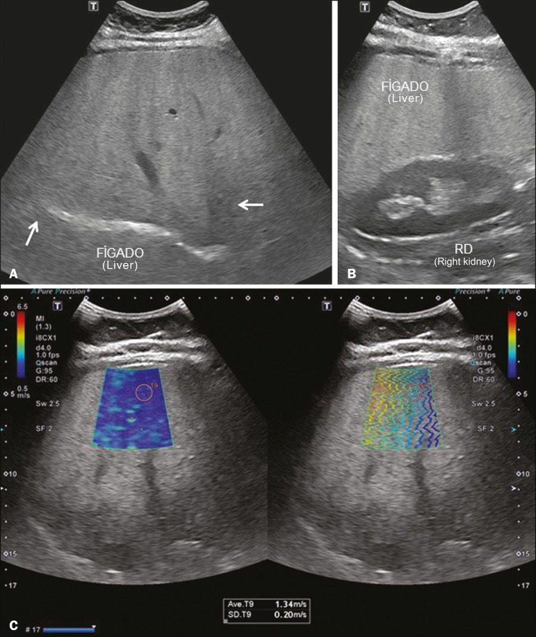
Hepatic steatosis, or fatty liver disease, occurs due to the accumulation of lipids in hepatocytes. When it becomes chronic, lobular inflammation develops and the disease can evolve to hepatic fibrosis, liver cirrhosis, or hepatocellular carcinoma. Early diagnosis is desirable because patients diagnosed in the early stage of the disease respond better to treatment. In the early stages of fatty liver disease, the physical examination is often unremarkable. Fatty liver disease and hepatic fibrosis can be diagnosed and monitored through laboratory tests, imaging, and biopsy. Among the imaging methods, ultrasound stands out as an effective means of diagnosing and following patients with liver disease. Ultrasound used in conjunction with elastography (ultrasound elastography) has recently shown great utility in the follow-up of such patients. Ultrasound elastography studies the degree of deformation (stiffness) of an organ or lesion, so that when there is hardening, fibrosis, or cirrhosis of the liver, those alterations are well demonstrated. In this review article, we discuss the application of the different types of ultrasound elastography for liver studies: transient elastography, point shear wave elastography, and two-dimensional shear wave elastography. Although magnetic resonance elastography may also be used in the analysis of liver fibrosis, it will not be addressed in this article.
肝脂肪变性,即脂肪肝病,是由于肝细胞内脂质蓄积所致。当它发展为慢性时,会出现小叶炎症,疾病可能会进展为肝纤维化、肝硬化或肝细胞癌。早期诊断很有必要,因为在疾病早期被诊断出的患者对治疗反应更好。在脂肪肝病的早期阶段,体格检查通常无明显异常。脂肪肝病和肝纤维化可以通过实验室检查、影像学检查和活检来诊断和监测。在影像学方法中,超声是诊断和随访肝病患者的有效手段。超声与弹性成像(超声弹性成像)结合使用,最近在这类患者的随访中显示出很大的实用性。超声弹性成像研究器官或病变的变形程度(硬度),因此当肝脏出现硬化、纤维化或肝硬化时,这些改变能得到很好的显示。在这篇综述文章中,我们讨论不同类型的超声弹性成像在肝脏研究中的应用:瞬时弹性成像、点剪切波弹性成像和二维剪切波弹性成像。虽然磁共振弹性成像也可用于肝纤维化分析,但本文将不涉及。