Yang Mengdi, Jiang Zhiyuan, Yao Guangyu, Wang Zhiyu, Sun Jing, Qin Huanlong, Zhao Hui
Department of Internal Oncology, Shanghai Jiao Tong University Affiliated Sixth People's Hospital, Shanghai, China.
Department of Gastrointestinal Surgery, Shanghai Tenth People's Hospital Affiliated With Tongji University, Shanghai, China.
Front Oncol. 2020 Apr 7;10:380. doi: 10.3389/fonc.2020.00380. eCollection 2020.
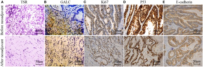
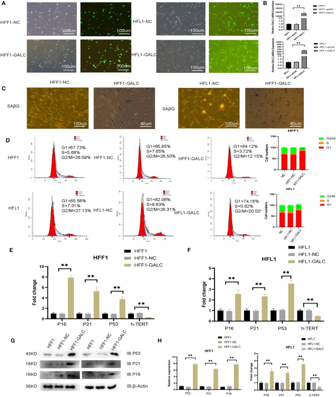
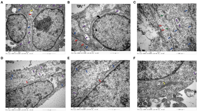
Colorectal cancer (CRC)-associated senescent fibroblasts may play a crucial role in tumor progression, but the mechanism remains unclear. In order to solve this complicated problem, we randomly collected 16 patients with CRC, who had been treated with oxaliplatin and capecitabine (XELOX). Hematoxylin-eosin (HE) staining revealed that the tumor-stroma ratio (TSR) of CRC was affected by XELOX treatment. Immunohistochemistry (IHC) and senescence-associated β-galactosidase (SAβG) staining were used to verify a stable model of senescent fibroblasts. IHC analysis showed that high expression levels of galactosylceramidase (GALC) and significant senescence-associated β-galactosidase (SAβG) staining were associated with CRC patient survival. We observed that fibroblasts overexpressing GALC underwent cell cycle arrest. Changes in cell morphology and cell cycle characteristics were accompanied by the upregulation of the , and gene, and the downregulation of expression. In a co-culture system, fibroblasts overexpressing GALC significantly increased the proliferation of CRC cells. Transmission electron microscopy (TEM) analysis confirmed that GALC overexpression fibroblasts co-cultured with CRC caused changes in CRC cell morphology. The aging fibroblast co-culture group (70%) had a higher migration ability. experiments and transcriptomics analysis were performed to verify the effect of senescent fibroblasts on tumor formation and to identify the potential mechanisms for the above results. We found that a high expression of ATF3 was related to good survival rates. However, a high expression of KIAA0907 was bad for survival rates ( < 0.05). The knockdown of ATF3 can promote cell proliferation, migration, and clonogenic assays, while downregulation of KIAA0907 inhibits cell proliferation, migration, and clonogenic assays. The results demonstrate that senescent fibroblasts with a high level of GALC regulated several aspects of the tumor growth process, including migration and invasion.
结直肠癌(CRC)相关的衰老成纤维细胞可能在肿瘤进展中起关键作用,但其机制仍不清楚。为了解决这个复杂的问题,我们随机收集了16例接受过奥沙利铂和卡培他滨(XELOX)治疗的CRC患者。苏木精-伊红(HE)染色显示,XELOX治疗会影响CRC的肿瘤-基质比(TSR)。免疫组织化学(IHC)和衰老相关β-半乳糖苷酶(SAβG)染色用于验证衰老成纤维细胞的稳定模型。IHC分析表明,半乳糖神经酰胺酶(GALC)的高表达水平和显著的衰老相关β-半乳糖苷酶(SAβG)染色与CRC患者的生存率相关。我们观察到过表达GALC的成纤维细胞会发生细胞周期停滞。细胞形态和细胞周期特征的变化伴随着 、 和 基因的上调以及 表达的下调。在共培养系统中,过表达GALC的成纤维细胞显著增加了CRC细胞的增殖。透射电子显微镜(TEM)分析证实,与CRC共培养的过表达GALC成纤维细胞导致CRC细胞形态发生变化。衰老成纤维细胞共培养组(70%)具有更高的迁移能力。进行了 实验和转录组学分析,以验证衰老成纤维细胞对肿瘤形成的影响,并确定上述结果的潜在机制。我们发现,ATF3的高表达与良好的生存率相关。然而,KIAA0907的高表达对生存率不利( < 0.05)。敲低ATF3可促进细胞增殖、迁移和克隆形成试验,而下调KIAA0907则抑制细胞增殖、迁移和克隆形成试验。结果表明,高水平GALC的衰老成纤维细胞调节了肿瘤生长过程的多个方面,包括迁移和侵袭。