González-Gutiérrez Alejandra G, Verdín Jorge, Rodríguez-Garay Benjamín
Unidad de Biotecnología Vegetal, CIATEJ, Centro de Investigación y Asistencia en Tecnología y Diseño del Estado de Jalisco, A.C., Zapopan, Mexico.
Unidad de Biotecnología Industrial, CIATEJ, Centro de Investigación y Asistencia en Tecnología y Diseño del Estado de Jalisco, A.C., Zapopan, Mexico.
Front Plant Sci. 2020 Apr 9;11:384. doi: 10.3389/fpls.2020.00384. eCollection 2020.
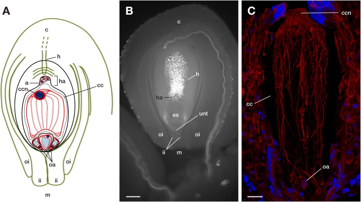
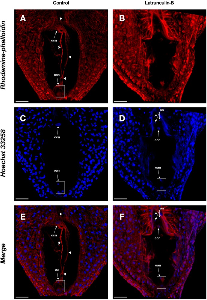
During plant sexual reproduction, F-actin takes part in the elongation of the pollen tube and the movement of sperm cells along with it. Moreover, F-actin is involved in the transport of sperm cells throughout the embryo sac when double fertilization occurs. Different techniques for analysis of F-actin in plant cells have been developed: from classical actin-immunolocalization in fixed tissues to genetically tagged actin with fluorescent proteins for live imaging of cells. Despite the implementation of live cell imaging tools, fixed plant tissue methods for cytoskeletal studies remain an essential tool for genetically intractable systems. Also, most of the work on live imaging of the cytoskeleton has been conducted on cells located on the plant's surface, such as epidermal cells, trichomes, and root hairs. In cells situated in the plant's interior, especially those from plant species with thicker organ systems, it is necessary to utilize conventional sectioning and permeabilization methods to allow the label access to the cytoskeleton. Studies about the role of F-actin cytoskeleton during double fertilization in plants with crassinucellate ovules (e.g., , and ) remain scarce due to the difficulties to access the female gametophyte. Here, we have developed a straightforward method for analysis of F-actin in the female gametophyte of different Agavoideae sub-family species. The procedure includes the fixation of whole ovules with formaldehyde, followed by membrane permeabilization with cold acetone, a prolonged staining step with rhodamine-phalloidin, and Hoechst 33342 as a counterstain and two final steps of dehydration of samples in increasing-concentration series of cold isopropanol and clarification of tissues with methyl salicylate. This technique allows the analysis of a large number of samples in a short period, cell positioning relative to neighbor cells is maintained, and, with the help of a confocal microscope, reconstruction of a single 3D image of F-actin structures into the embryo sac can be obtained.
在植物有性生殖过程中,丝状肌动蛋白(F-肌动蛋白)参与花粉管的伸长以及精细胞随之进行的移动。此外,在双受精发生时,F-肌动蛋白还参与精细胞在整个胚囊中的运输。人们已经开发出多种用于分析植物细胞中F-肌动蛋白的技术:从固定组织中的经典肌动蛋白免疫定位,到利用荧光蛋白进行基因标记的肌动蛋白以对细胞进行实时成像。尽管有了活细胞成像工具,但用于细胞骨架研究的固定植物组织方法仍是研究遗传难处理系统的重要工具。而且,大多数关于细胞骨架实时成像的工作都是在植物表面的细胞上进行的,如表皮细胞、毛状体和根毛。对于位于植物内部的细胞,尤其是那些来自器官系统较厚的植物物种的细胞,有必要采用传统的切片和通透方法,以使标记物能够接触到细胞骨架。由于难以获取雌配子体,关于具厚珠心胚珠的植物(如 、 和 )双受精过程中F-肌动蛋白细胞骨架作用的研究仍然很少。在此,我们开发了一种直接的方法来分析不同龙舌兰亚科物种雌配子体中的F-肌动蛋白。该程序包括用甲醛固定整个胚珠,随后用冷丙酮进行膜通透处理,用罗丹明-鬼笔环肽进行长时间染色步骤,并用Hoechst 33342作为复染剂,以及最后两个步骤,即先用浓度递增的冷异丙醇系列对样品进行脱水,再用冬青油对组织进行透明处理。该技术能够在短时间内分析大量样品,保持细胞相对于相邻细胞的位置,并且借助共聚焦显微镜,可以获得F-肌动蛋白结构在胚囊内的单个三维图像重建。