School of Engineering and Materials Science, Queen Mary University of London, Mile End Road, E1 4NS, London, UK.
Sci Rep. 2019 Mar 1;9(1):3241. doi: 10.1038/s41598-019-40092-w.
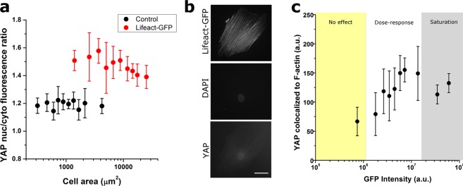
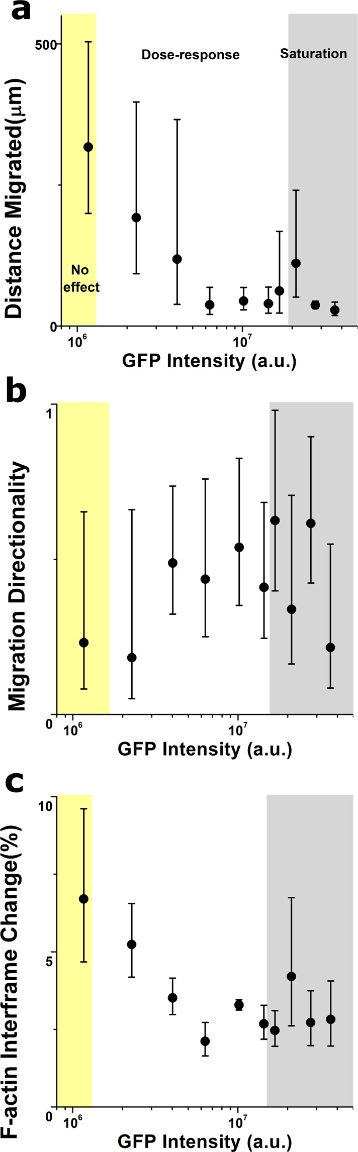
Live-imaging techniques are at the forefront of biology research to explore behaviour and function from sub-cellular to whole organism scales. These methods rely on intracellular fluorescent probes to label specific proteins, which are commonly assumed to only introduce artefacts at concentrations far-exceeding routine use. Lifeact, a small peptide with affinity for actin microfilaments has become a gold standard in live cell imaging of the cytoskeleton. Nevertheless, recent reports have raised concerns on Lifeact-associated artefacts at the molecular and whole organism level. We show here that Lifeact induces dose-response artefacts at the cellular level, impacting stress fibre dynamics and actin cytoskeleton architecture. These effects extend to the microtubule and intermediate filament networks as well as the nucleus, and ultimately lead to altered subcellular localization of YAP, reduced cell migration and abnormal mechanical properties. Our results suggest that reduced binding of cofilin to actin filaments may be the underlying cause of the observed Lifeact-induced cellular artefacts.
活体成像技术是生物学研究的前沿领域,可从亚细胞到整个生物体的尺度探索行为和功能。这些方法依赖于细胞内荧光探针来标记特定的蛋白质,通常认为这些蛋白质只有在浓度远远超过常规使用的情况下才会引入伪影。Lifeact 是一种与肌动蛋白微丝亲和力强的小肽,已成为活细胞骨架成像的金标准。然而,最近的报道引起了人们对 Lifeact 相关分子和整个生物体水平上的伪影的关注。我们在这里表明,Lifeact 在细胞水平上诱导剂量反应伪影,影响应力纤维动力学和肌动蛋白细胞骨架结构。这些影响扩展到微管和中间丝网络以及细胞核,并最终导致 YAP 的亚细胞定位改变、细胞迁移减少和异常的力学特性。我们的结果表明,肌动蛋白丝结合的 cofilin 减少可能是观察到的 Lifeact 诱导的细胞伪影的根本原因。