• 文献检索
  • 文档翻译
  • 深度研究
  • 学术资讯
  • Suppr Zotero 插件Zotero 插件
  • 邀请有礼
  • 套餐&价格
  • 历史记录
应用&插件
Suppr Zotero 插件Zotero 插件浏览器插件Mac 客户端Windows 客户端微信小程序
定价
高级版会员购买积分包购买API积分包
服务
文献检索文档翻译深度研究API 文档MCP 服务
关于我们
关于 Suppr公司介绍联系我们用户协议隐私条款
关注我们

Suppr 超能文献

核心技术专利:CN118964589B侵权必究
粤ICP备2023148730 号-1Suppr @ 2026

文献检索

告别复杂PubMed语法,用中文像聊天一样搜索,搜遍4000万医学文献。AI智能推荐,让科研检索更轻松。

立即免费搜索

文件翻译

保留排版,准确专业,支持PDF/Word/PPT等文件格式,支持 12+语言互译。

免费翻译文档

深度研究

AI帮你快速写综述,25分钟生成高质量综述,智能提取关键信息,辅助科研写作。

立即免费体验

一种基于共聚焦显微镜和厚切片的简单方法可识别骺板软骨细胞中的七个亚时相。

A simple method based on confocal microscopy and thick sections recognizes seven subphases in growth plate chondrocytes.

机构信息

Division of Pediatrics, Department of Medicine, Faculty of Medicine, University of Oviedo, CP 33006, Oviedo, Asturias, Spain.

Fundacion para la Investigación Sanitaria del Principado de Asturias (FINBA), Oviedo, Spain.

出版信息

Sci Rep. 2020 Apr 24;10(1):6935. doi: 10.1038/s41598-020-63978-6.

DOI:10.1038/s41598-020-63978-6
PMID:32332842
原文链接:https://pmc.ncbi.nlm.nih.gov/articles/PMC7181680/
Abstract

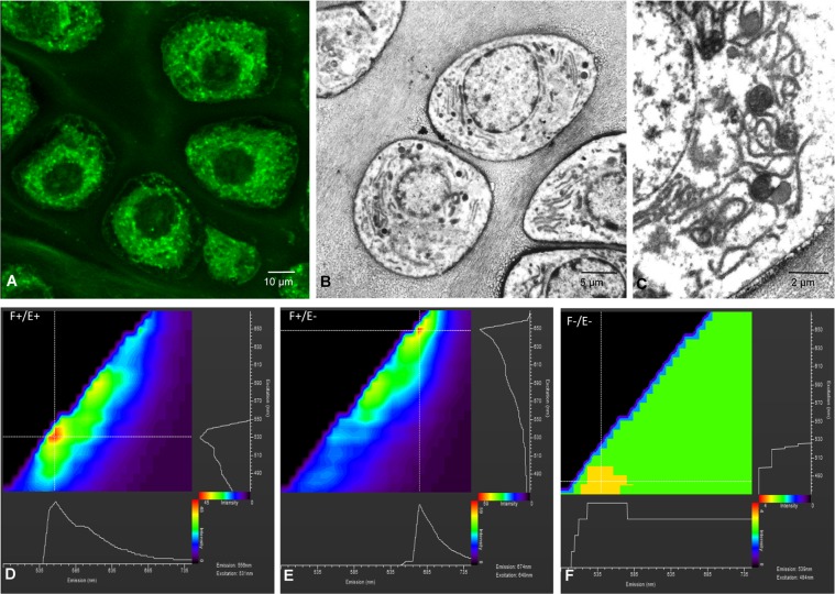
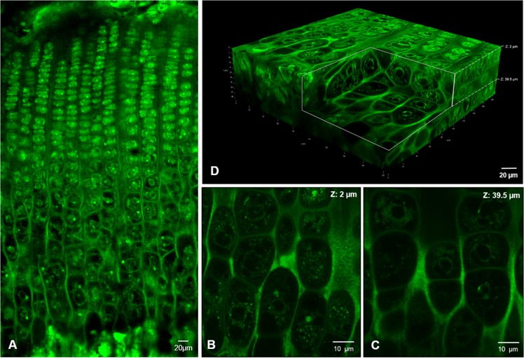
This manuscript reports a novel procedure to imaging growth plate chondrocytes by using confocal microscopy. The method is based on fixed undecalcified bone samples, in-block staining with eosin, epoxy resin embedding and grinding to obtain thick sections. It is simple, inexpensive and provides three-dimensional images of entire chondrocytes inside their native lacunae. Quantitative analysis of volume, shape and cytoplasm density of chondrocytes at different strata of the growth plate allowed to objectively grade chondrocytes of the growth plate in seven different clusters. These seven categories of chondrocytes were subsequently evaluated by immunohistochemistry of some well-defined molecular landmarks of chondrocyte differentiation. Furthermore, immunohistochemical analysis of proteins responsible for ionic changes and water transport allowing chondrocyte swelling during hypertrophy was also performed. Results obtained indicate that four subphases can be defined in the pre-hypertrophic zone and three subphases in the hypertrophic zone, a fact that raises that chondrocytes of the growth plate are less homogeneous than usually considered when different zones are defined according to subjective cell morphological criteria. Results in the present study provide a technological innovation and gives new insights into the complexity of the process of chondrocyte differentiation in the growth plate.

摘要

这篇手稿报告了一种通过共聚焦显微镜对生长板软骨细胞进行成像的新方法。该方法基于固定的未脱钙骨样本,采用伊红、环氧树脂包埋和研磨进行块内染色,以获得厚切片。它简单、廉价,并提供了整个软骨细胞在其天然腔中的三维图像。对生长板不同层的软骨细胞的体积、形状和细胞质密度进行定量分析,能够客观地将生长板软骨细胞分为七个不同的簇。然后,通过对一些明确的软骨细胞分化分子标志物的免疫组织化学分析,对这 7 类软骨细胞进行评估。此外,还对负责离子变化和水运输的蛋白质进行了免疫组织化学分析,这些蛋白质允许软骨细胞在肥大过程中肿胀。研究结果表明,在预备肥大区可以定义四个亚区,在肥大区可以定义三个亚区,这一事实表明,与根据主观细胞形态学标准定义不同区域时相比,生长板中的软骨细胞的同质性较差。本研究的结果提供了技术创新,并深入了解了生长板中软骨细胞分化过程的复杂性。

https://cdn.ncbi.nlm.nih.gov/pmc/blobs/baea/7181680/efd8deb97ab0/41598_2020_63978_Fig5_HTML.jpg
https://cdn.ncbi.nlm.nih.gov/pmc/blobs/baea/7181680/2b5f2b9ddce1/41598_2020_63978_Fig1_HTML.jpg
https://cdn.ncbi.nlm.nih.gov/pmc/blobs/baea/7181680/90cfeb2d3d2d/41598_2020_63978_Fig2_HTML.jpg
https://cdn.ncbi.nlm.nih.gov/pmc/blobs/baea/7181680/33fc73c976fc/41598_2020_63978_Fig3_HTML.jpg
https://cdn.ncbi.nlm.nih.gov/pmc/blobs/baea/7181680/736fe46f9f13/41598_2020_63978_Fig4_HTML.jpg
https://cdn.ncbi.nlm.nih.gov/pmc/blobs/baea/7181680/efd8deb97ab0/41598_2020_63978_Fig5_HTML.jpg

相似文献

1
A simple method based on confocal microscopy and thick sections recognizes seven subphases in growth plate chondrocytes.一种基于共聚焦显微镜和厚切片的简单方法可识别骺板软骨细胞中的七个亚时相。
Sci Rep. 2020 Apr 24;10(1):6935. doi: 10.1038/s41598-020-63978-6.
2
Microtubules are potential regulators of growth-plate chondrocyte differentiation and hypertrophy.微管是生长板软骨细胞分化和肥大的潜在调节因子。
Bone. 1999 Oct;25(4):405-12. doi: 10.1016/s8756-3282(99)00187-8.
3
The osmotic sensitivity of rat growth plate chondrocytes in situ; clarifying the mechanisms of hypertrophy.大鼠生长板软骨细胞原位的渗透敏感性;阐明肥大机制。
J Cell Physiol. 2008 Mar;214(3):621-9. doi: 10.1002/jcp.21249.
4
Three-dimensional in situ zonal morphology of viable growth plate chondrocytes: a confocal microscopy study.三维共聚焦显微镜观察存活生长板软骨细胞的区域性形态。
J Orthop Res. 2011 May;29(5):710-7. doi: 10.1002/jor.21294. Epub 2010 Nov 30.
5
Primary culture of rat growth plate chondrocytes: an in vitro model of growth plate histotype, matrix vesicle biogenesis and mineralization.大鼠生长板软骨细胞的原代培养:生长板组织类型、基质小泡生物发生及矿化的体外模型
Bone. 2004 Jun;34(6):961-70. doi: 10.1016/j.bone.2004.02.010.
6
The clustering and morphology of chondrocytes in normal and mildly degenerate human femoral head cartilage studied by confocal laser scanning microscopy.通过共聚焦激光扫描显微镜研究正常和轻度退变的人股骨头软骨中软骨细胞的聚集和形态。
J Anat. 2018 Apr;232(4):686-698. doi: 10.1111/joa.12768. Epub 2017 Dec 28.
7
Tissue and cellular morphological changes in growth plate explants under compression.压缩条件下生长板外植体的组织和细胞形态变化。
J Biomech. 2010 Sep 17;43(13):2582-8. doi: 10.1016/j.jbiomech.2010.05.010.
8
mir-374-5p, mir-379-5p, and mir-503-5p Regulate Proliferation and Hypertrophic Differentiation of Growth Plate Chondrocytes in Male Rats.miR-374-5p、miR-379-5p 和 miR-503-5p 调控雄性大鼠生长板软骨细胞的增殖和肥大分化。
Endocrinology. 2018 Mar 1;159(3):1469-1478. doi: 10.1210/en.2017-00780.
9
Progression and recapitulation of the chondrocyte differentiation program: cartilage matrix protein is a marker for cartilage maturation.软骨细胞分化程序的进展与重现:软骨基质蛋白是软骨成熟的标志物。
Dev Biol. 1995 Nov;172(1):293-306. doi: 10.1006/dbio.1995.0024.
10
Sox5 and Sox6 are needed to develop and maintain source, columnar, and hypertrophic chondrocytes in the cartilage growth plate.Sox5和Sox6是软骨生长板中发育和维持源细胞、柱状细胞和肥大软骨细胞所必需的。
J Cell Biol. 2004 Mar 1;164(5):747-58. doi: 10.1083/jcb.200312045.

引用本文的文献

1
In-situ microscopy and digital image correlation to study the mechanical characteristics of polymer-based materials.利用原位显微镜和数字图像相关技术研究聚合物基材料的力学特性。
Discov Mater. 2025;5(1):41. doi: 10.1007/s43939-025-00208-8. Epub 2025 Feb 18.
2
Roles of Sodium Hydrogen Exchanger (NHE1) and Anion Exchanger (AE2) across Chondrocytes Plasma Membrane during Longitudinal Bone Growth.钠氢交换体(NHE1)和阴离子交换体(AE2)在纵向骨生长过程中跨软骨细胞质膜的作用。
Membranes (Basel). 2022 Jul 14;12(7):707. doi: 10.3390/membranes12070707.
3
Cellular and Molecular Alterations Underlying Abnormal Bone Growth in X-Linked Hypophosphatemia.
X 连锁低磷血症异常骨生长的细胞和分子改变。
Int J Mol Sci. 2022 Jan 15;23(2):934. doi: 10.3390/ijms23020934.
4
Effect of Ivermectin and Atorvastatin on Nuclear Localization of Importin Alpha and Drug Target Expression Profiling in Host Cells from Nasopharyngeal Swabs of SARS-CoV-2- Positive Patients.伊维菌素和阿托伐他汀对 SARS-CoV-2 阳性患者鼻咽拭子中宿主细胞内 Importin Alpha 核定位及药物靶标表达谱的影响。
Viruses. 2021 Oct 15;13(10):2084. doi: 10.3390/v13102084.
5
Enlightenment of Growth Plate Regeneration Based on Cartilage Repair Theory: A Review.基于软骨修复理论的生长板再生研究进展:综述
Front Bioeng Biotechnol. 2021 Jun 3;9:654087. doi: 10.3389/fbioe.2021.654087. eCollection 2021.
6
The Formation of the Epiphyseal Bone Plate Occurs via Combined Endochondral and Intramembranous-Like Ossification.骺板的形成是通过软骨内骨化和膜内骨化相结合的方式进行的。
Int J Mol Sci. 2021 Jan 18;22(2):900. doi: 10.3390/ijms22020900.
7
Innovative Three-Dimensional Microscopic Analysis of Uremic Growth Plate Discloses Alterations in the Process of Chondrocyte Hypertrophy: Effects of Growth Hormone Treatment.创新性三维微观分析尿毒症生长板揭示了软骨细胞肥大过程中的变化:生长激素治疗的影响。
Int J Mol Sci. 2020 Jun 25;21(12):4519. doi: 10.3390/ijms21124519.