• 文献检索
  • 文档翻译
  • 深度研究
  • 学术资讯
  • Suppr Zotero 插件Zotero 插件
  • 邀请有礼
  • 套餐&价格
  • 历史记录
应用&插件
Suppr Zotero 插件Zotero 插件浏览器插件Mac 客户端Windows 客户端微信小程序
定价
高级版会员购买积分包购买API积分包
服务
文献检索文档翻译深度研究API 文档MCP 服务
关于我们
关于 Suppr公司介绍联系我们用户协议隐私条款
关注我们

Suppr 超能文献

核心技术专利:CN118964589B侵权必究
粤ICP备2023148730 号-1Suppr @ 2026

文献检索

告别复杂PubMed语法,用中文像聊天一样搜索,搜遍4000万医学文献。AI智能推荐,让科研检索更轻松。

立即免费搜索

文件翻译

保留排版,准确专业,支持PDF/Word/PPT等文件格式,支持 12+语言互译。

免费翻译文档

深度研究

AI帮你快速写综述,25分钟生成高质量综述,智能提取关键信息,辅助科研写作。

立即免费体验

对比增强超声检查(CEUS)用于检测新型冠状病毒肺炎(COVID-19)严重感染病例的腹部微循环障碍:初步经验

Contrast enhanced ultrasonography (CEUS) to detect abdominal microcirculatory disorders in severe cases of COVID-19 infection: First experience.

作者信息

Jung E M, Stroszczynski C, Jung F

机构信息

Institute of Diagnostic Radiology, University Hospital, Regensburg, Germany.

Interdisciplinary Ultrasound Department, University Hospital, Regensburg, Germany.

出版信息

Clin Hemorheol Microcirc. 2020;74(4):353-361. doi: 10.3233/CH-209003.

DOI:10.3233/CH-209003
PMID:32333581
原文链接:https://pmc.ncbi.nlm.nih.gov/articles/PMC7369109/
Abstract

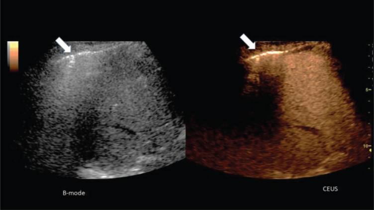
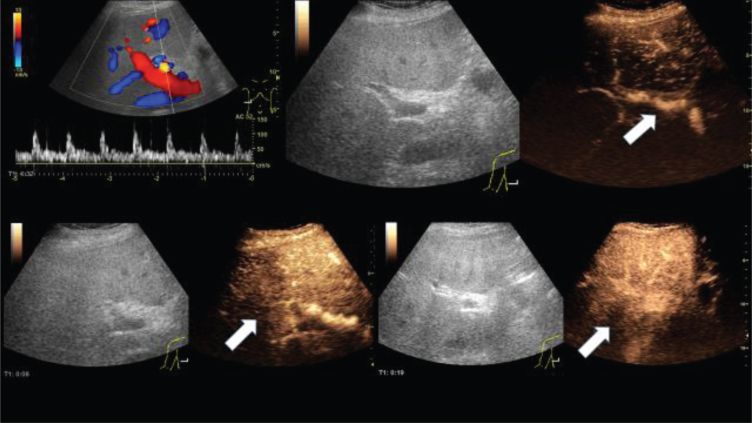
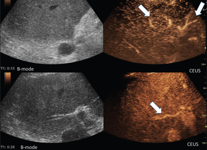
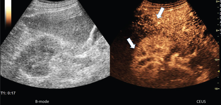
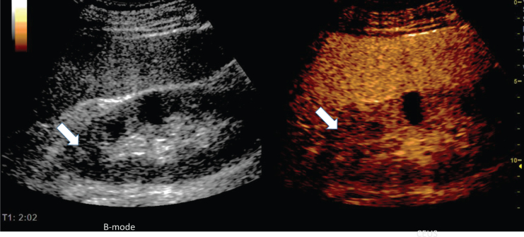
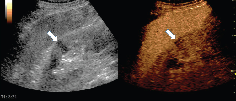
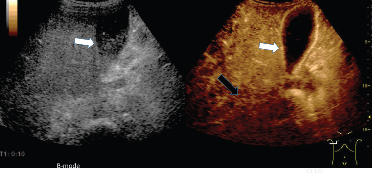
In the hands of experienced examiners, the contrast enhanced sonography (CEUS) offers the possibility to analyze dynamic microcirculatory disturbances in real time dynamically without any risk for kidneys and thyroid gland even in severe progressing disease bedside. Based on severe COVID-19 infections, first experiences with abdominal CEUS examinations are presented. In the stage of an imminent organ failure with significantly reduced kidney and liver function, CEUS can be used to show a narrowing of the organ-supplying arteries, as well as a delayed capillary filling of vessels near the capsule, a regional reduced parenchymal perfusion or an inflammatory hyperemia with capillary hypercirculation. It is possible to quickly rule out organ infarction and to dynamically record the mesenteric arterial and venous blood flow.

摘要

在经验丰富的检查者手中,超声造影(CEUS)能够实时动态分析动态微循环紊乱,即使在床边严重进展性疾病的情况下,对肾脏和甲状腺也没有任何风险。基于严重的COVID-19感染,介绍了腹部CEUS检查的初步经验。在即将发生器官衰竭且肾脏和肝功能显著降低的阶段,CEUS可用于显示器官供血动脉变窄,以及包膜附近血管的毛细血管充盈延迟、局部实质灌注减少或伴有毛细血管过度循环的炎症性充血。可以快速排除器官梗死,并动态记录肠系膜动脉和静脉血流。

https://cdn.ncbi.nlm.nih.gov/pmc/blobs/463e/7369109/461b9c5b0ade/ch-74-ch209003-g007.jpg
https://cdn.ncbi.nlm.nih.gov/pmc/blobs/463e/7369109/0910c19da026/ch-74-ch209003-g001.jpg
https://cdn.ncbi.nlm.nih.gov/pmc/blobs/463e/7369109/8b0ea1698ae7/ch-74-ch209003-g002.jpg
https://cdn.ncbi.nlm.nih.gov/pmc/blobs/463e/7369109/349590cadf31/ch-74-ch209003-g003.jpg
https://cdn.ncbi.nlm.nih.gov/pmc/blobs/463e/7369109/3672059b8471/ch-74-ch209003-g004.jpg
https://cdn.ncbi.nlm.nih.gov/pmc/blobs/463e/7369109/7231a32d71eb/ch-74-ch209003-g005.jpg
https://cdn.ncbi.nlm.nih.gov/pmc/blobs/463e/7369109/2b81c003e9c5/ch-74-ch209003-g006.jpg
https://cdn.ncbi.nlm.nih.gov/pmc/blobs/463e/7369109/461b9c5b0ade/ch-74-ch209003-g007.jpg

相似文献

1
Contrast enhanced ultrasonography (CEUS) to detect abdominal microcirculatory disorders in severe cases of COVID-19 infection: First experience.对比增强超声检查(CEUS)用于检测新型冠状病毒肺炎(COVID-19)严重感染病例的腹部微循环障碍:初步经验
Clin Hemorheol Microcirc. 2020;74(4):353-361. doi: 10.3233/CH-209003.
2
Contrast enhanced ultrasound (CEUS) to assess pleural pulmonal changes in severe COVID-19 infection: First results.超声造影(CEUS)评估重症 COVID-19 感染患者的胸膜肺改变:初步结果。
Clin Hemorheol Microcirc. 2020;75(1):19-26. doi: 10.3233/CH-209005.
3
[Pulmonary Sonography in Patients With Suspected COVID-19 - Step-by-Step].[疑似新型冠状病毒肺炎患者的肺部超声检查——分步指南]
Dtsch Med Wochenschr. 2020 Jul;145(15):1100-1104. doi: 10.1055/a-1185-6885. Epub 2020 Jul 30.
4
Contrast-enhanced ultrasound (CEUS) of the lung reveals multiple areas of microthrombi in a COVID-19 patient.一名新冠肺炎患者的肺部对比增强超声(CEUS)显示多个微血栓区域。
Intensive Care Med. 2020 Aug;46(8):1660-1662. doi: 10.1007/s00134-020-06085-4. Epub 2020 May 18.
5
Lung ultrasound findings in patients with COVID-19 pneumonia.新型冠状病毒肺炎患者的肺部超声检查结果
Crit Care. 2020 Apr 28;24(1):174. doi: 10.1186/s13054-020-02876-9.
6
Thoracic ultrasound and SARS-COVID-19: a pictorial essay.胸部超声与新型冠状病毒肺炎:图文并茂的文章
J Ultrasound. 2020 Jun;23(2):217-221. doi: 10.1007/s40477-020-00458-7. Epub 2020 Apr 16.
7
Ultrasonography and SARS-CoV 2 infection: a review of what we know and do not yet know.超声检查与2019冠状病毒病感染:我们已知与未知情况的综述
Med Ultrason. 2020 May 11;22(2):129-132. doi: 10.11152/mu-2612.
8
Lung Ultrasound in COVID-19.新型冠状病毒肺炎中的肺部超声
Indian Pediatr. 2020 Aug 15;57(8):774. doi: 10.1007/s13312-020-1942-3. Epub 2020 Jul 24.
9
Lung ultrasound in the frontline diagnosis of COVID-19 infection.肺部超声在新型冠状病毒肺炎感染的一线诊断中的应用
Med Clin (Barc). 2020 Sep 11;155(5):232. doi: 10.1016/j.medcli.2020.06.001. Epub 2020 Jun 12.
10
Is There a Role for Lung Ultrasound During the COVID-19 Pandemic?在新冠疫情期间,肺部超声能发挥作用吗?
J Ultrasound Med. 2020 Jul;39(7):1459-1462. doi: 10.1002/jum.15284. Epub 2020 Apr 7.

引用本文的文献

1
Contrast-Enhanced Intraoperative Ultrasound Shows Excellent Performance in Improving Intraoperative Decision-Making.术中超声造影在改善术中决策方面表现出色。
Life (Basel). 2024 Sep 22;14(9):1199. doi: 10.3390/life14091199.
2
A Blood Supply Pathophysiological Microcirculatory Mechanism for Long COVID.长期新冠的一种血液供应病理生理微循环机制
Life (Basel). 2024 Aug 28;14(9):1076. doi: 10.3390/life14091076.
3
Morphology and Function of Red Blood Cells in COVID-19 Patients: Current Overview 2023.2023年新冠病毒感染患者红细胞的形态与功能:当前概述

本文引用的文献

1
[Comparison of clinical and pathological features between severe acute respiratory syndrome and coronavirus disease 2019].严重急性呼吸综合征与2019冠状病毒病的临床和病理特征比较
Zhonghua Jie He He Hu Xi Za Zhi. 2020 Jun 12;43(6):496-502. doi: 10.3760/cma.j.cn112147-20200311-00312.
2
Early Clinical and CT Manifestations of Coronavirus Disease 2019 (COVID-19) Pneumonia.新型冠状病毒肺炎(COVID-19)肺炎的早期临床和 CT 表现。
AJR Am J Roentgenol. 2020 Aug;215(2):338-343. doi: 10.2214/AJR.20.22961. Epub 2020 Mar 17.
3
Findings of lung ultrasonography of novel corona virus pneumonia during the 2019-2020 epidemic.
Life (Basel). 2024 Apr 1;14(4):460. doi: 10.3390/life14040460.
4
Abdominal Imaging Findings in Patients with COVID-19 Part 2: Solid Organs.新型冠状病毒肺炎患者的腹部影像学表现 第2部分:实体器官
Middle East J Dig Dis. 2022 Oct;14(4):373-381. doi: 10.34172/mejdd.2022.298. Epub 2022 Oct 30.
5
Potential Benefits of Omega-3 Polyunsaturated Fatty Acids (N3PUFAs) on Cardiovascular Health Associated with COVID-19: An Update for 2023.欧米伽-3多不饱和脂肪酸(N3PUFAs)对与COVID-19相关的心血管健康的潜在益处:2023年最新情况
Metabolites. 2023 May 5;13(5):630. doi: 10.3390/metabo13050630.
6
Findings of Abdominal Imaging in Patients with COVID-19 - Part 1: Hollow Organs.新型冠状病毒肺炎患者腹部影像学表现——第1部分:中空脏器
Middle East J Dig Dis. 2022 Jul;14(3):278-286. doi: 10.34172/mejdd.2022.284. Epub 2022 Jul 30.
7
Probing Red Blood Cell Membrane Microviscosity Using Fluorescence Anisotropy Decay Curves of the Lipophilic Dye PKH26.利用亲脂性染料 PKH26 的荧光各向异性衰减曲线探测红细胞膜微观粘度。
Int J Mol Sci. 2022 Dec 12;23(24):15767. doi: 10.3390/ijms232415767.
8
Blood hyperviscosity in acute and recent COVID-19 infection.急性和近期 COVID-19 感染中的血液高黏度。
Clin Hemorheol Microcirc. 2022;82(2):149-155. doi: 10.3233/CH-221429.
9
The Value of Contrast-Enhanced Ultrasound (CEUS) in the Detection of Perfusion Disturbances in Abdominal Wall Hernias Compared with Surgical and Histological Assessment.与手术及组织学评估相比,对比增强超声(CEUS)在检测腹壁疝灌注异常中的价值
Diagnostics (Basel). 2022 Feb 1;12(2):370. doi: 10.3390/diagnostics12020370.
10
Vascular endotheliitis associated with infections: Its pathogenetic role and therapeutic implication.感染相关血管内皮炎:发病机制作用及其治疗意义。
Biochem Pharmacol. 2022 Mar;197:114909. doi: 10.1016/j.bcp.2022.114909. Epub 2022 Jan 10.
2019 - 2020年疫情期间新型冠状病毒肺炎的肺部超声检查结果
Intensive Care Med. 2020 May;46(5):849-850. doi: 10.1007/s00134-020-05996-6. Epub 2020 Mar 12.
4
Corona Virus International Public Health Emergencies: Implications for Radiology Management.冠状病毒国际公共卫生突发事件:对放射科管理的影响。
Acad Radiol. 2020 Apr;27(4):463-467. doi: 10.1016/j.acra.2020.02.003. Epub 2020 Feb 26.
5
Clinical course and outcomes of critically ill patients with SARS-CoV-2 pneumonia in Wuhan, China: a single-centered, retrospective, observational study.中国武汉严重 COVID-19 患者的临床病程和结局:一项单中心、回顾性、观察性研究。
Lancet Respir Med. 2020 May;8(5):475-481. doi: 10.1016/S2213-2600(20)30079-5. Epub 2020 Feb 24.
6
Contrast-enhanced ultrasound (CEUS) of cystic renal lesions in comparison to CT and MRI in a multicenter setting.对比增强超声(CEUS)与 CT 和 MRI 在多中心环境下对囊性肾病变的比较。
Clin Hemorheol Microcirc. 2020;75(4):419-429. doi: 10.3233/CH-190764.
7
Vascular pattern and diagnostic accuracy of contrast-enhanced ultrasound (CEUS) in spleen alterations.对比增强超声(CEUS)在脾脏改变中的血管模式和诊断准确性。
Clin Hemorheol Microcirc. 2020;75(2):177-188. doi: 10.3233/CH-190758.
8
Comparison of the Diagnostic Performance of Contrast-enhanced Ultrasound with That of Contrast-enhanced Computed Tomography and Contrast-enhanced Magnetic Resonance Imaging in the Evaluation of Renal Masses: A Systematic Review and Meta-analysis.对比增强超声与对比增强计算机断层扫描和对比增强磁共振成像在评估肾肿块中的诊断性能比较:系统评价和荟萃分析。
Eur Urol Oncol. 2020 Aug;3(4):464-473. doi: 10.1016/j.euo.2019.08.013. Epub 2019 Sep 27.
9
Narrative review: clinical assessment of peripheral tissue perfusion in septic shock.叙述性综述:脓毒性休克时外周组织灌注的临床评估
Ann Intensive Care. 2019 Mar 13;9(1):37. doi: 10.1186/s13613-019-0511-1.
10
Influence of limited examination conditions on contrast-enhanced sonography for characterising liver lesions.有限检查条件对肝脏病变超声造影特征分析的影响
Clin Hemorheol Microcirc. 2019;71(2):267-276. doi: 10.3233/CH-189417.