Li Ka Shing Institute of Virology and Department of Medical Microbiology and Immunology, University of Alberta, Edmonton, Alberta, Canada.
Alberta Diabetes Institute, University of Alberta, Edmonton, Alberta, Canada.
PLoS Pathog. 2020 Apr 30;16(4):e1008515. doi: 10.1371/journal.ppat.1008515. eCollection 2020 Apr.
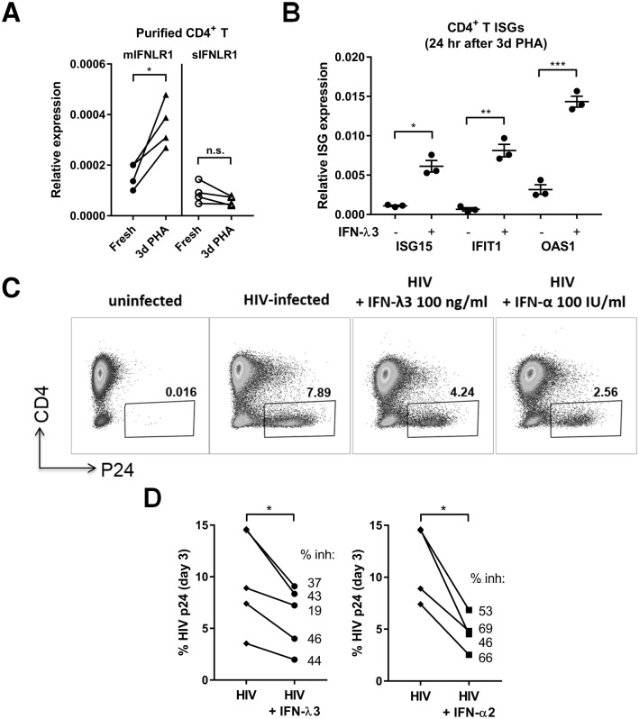
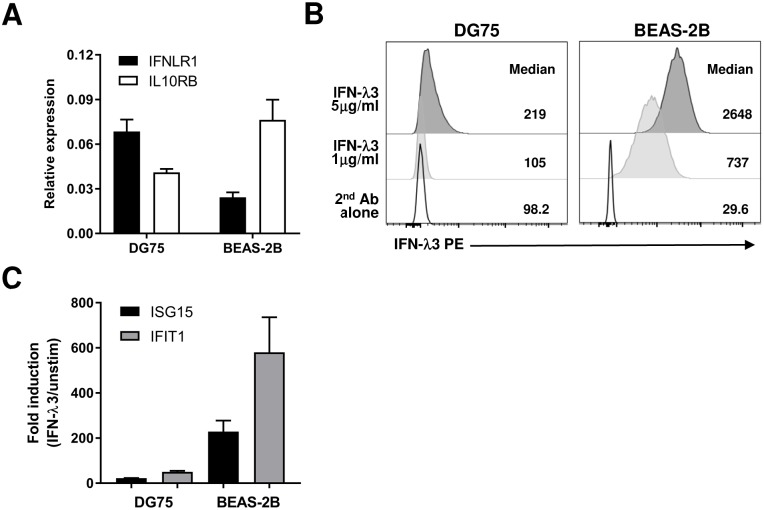
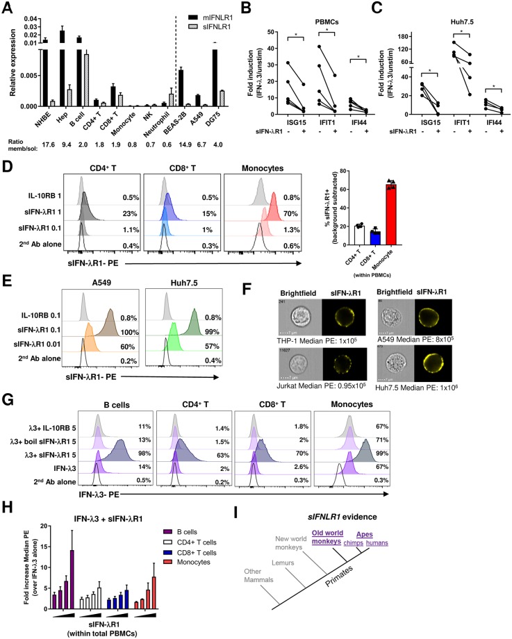
Type III interferons (IFN-lambdas(λ)) are important cytokines that inhibit viruses and modulate immune responses by acting through a unique IFN-λR1/IL-10RB heterodimeric receptor. Until now, the primary antiviral function of IFN-λs has been proposed to be at anatomical barrier sites. Here, we examine the regulation of IFN-λR1 expression and measure the downstream effects of IFN-λ3 stimulation in primary human blood immune cells, compared with lung or liver epithelial cells. IFN-λ3 directly bound and upregulated IFN-stimulated gene (ISG) expression in freshly purified human B cells and CD8+ T cells, but not monocytes, neutrophils, natural killer cells, and CD4+ T cells. Despite similar IFNLR1 transcript levels in B cells and lung epithelial cells, lung epithelial cells bound more IFN-λ3, which resulted in a 50-fold greater ISG induction when compared to B cells. The reduced response of B cells could be explained by higher expression of the soluble variant of IFN-λR1 (sIFN-λR1), which significantly reduced ISG induction when added with IFN-λ3 to peripheral blood mononuclear cells or liver epithelial cells. T-cell receptor stimulation potently, and specifically, upregulated membrane-bound IFNLR1 expression in CD4+ T cells, leading to greater antiviral gene induction, and inhibition of human immunodeficiency virus type 1 infection. Collectively, our data demonstrate IFN-λ3 directly interacts with the human adaptive immune system, unlike what has been previously shown in published mouse models, and that type III IFNs could be potentially utilized to suppress both mucosal and blood-borne viral infections.
III 型干扰素(IFN-λs)是重要的细胞因子,通过与独特的 IFN-λR1/IL-10RB 异二聚体受体相互作用,抑制病毒并调节免疫反应。到目前为止,IFN-λs 的主要抗病毒功能已被提议在解剖屏障部位发挥作用。在这里,我们研究了 IFN-λR1 表达的调节,并比较了肺或肝上皮细胞,测量了 IFN-λ3 刺激对原代人血免疫细胞的下游影响。IFN-λ3 直接结合并上调了新鲜纯化的人 B 细胞和 CD8+T 细胞中的 IFN 刺激基因(ISG)表达,但不结合单核细胞、中性粒细胞、自然杀伤细胞和 CD4+T 细胞。尽管 B 细胞和肺上皮细胞中的 IFNLR1 转录本水平相似,但肺上皮细胞结合了更多的 IFN-λ3,与 B 细胞相比,诱导的 ISG 表达增加了 50 倍。B 细胞的反应减少可以用可溶性 IFN-λR1(sIFN-λR1)的高表达来解释,当将 sIFN-λR1 与 IFN-λ3 一起添加到外周血单核细胞或肝上皮细胞中时,sIFN-λR1 显著降低了 ISG 诱导。T 细胞受体刺激强烈且特异性地上调了 CD4+T 细胞中膜结合 IFNLR1 的表达,导致更大的抗病毒基因诱导,并抑制了人类免疫缺陷病毒 1 型的感染。总的来说,我们的数据表明,IFN-λ3 直接与人类适应性免疫系统相互作用,与之前在已发表的小鼠模型中所显示的不同,并且 III 型 IFNs 可能被潜在地用于抑制粘膜和血源病毒感染。